Serum neuron specific enolase is increased in pediatric acute encephalitis syndrome
Article information
Abstract
Purpose
This study aimed to investigate whether serum neuron-specific enolase (NSE) was expressed in acute encephalitis syndrome (AES) that causes neuronal damage in children.
Methods
This prospective observational study was conducted in the pediatric neurology ward of Soetomo Hospital. Cases of AES with ages ranging from 1 month to 12 years were included. Cases that were categorized as simple and complex febrile seizures constituted the non-AES group. Blood was collected for the measurement of NSE within 24 hours of hemodynamic stabilization. The median NSE values of both groups were compared by using the Mann-Whitney U test. All statistical analyses were performed with SPSS version 12 for Windows.
Results
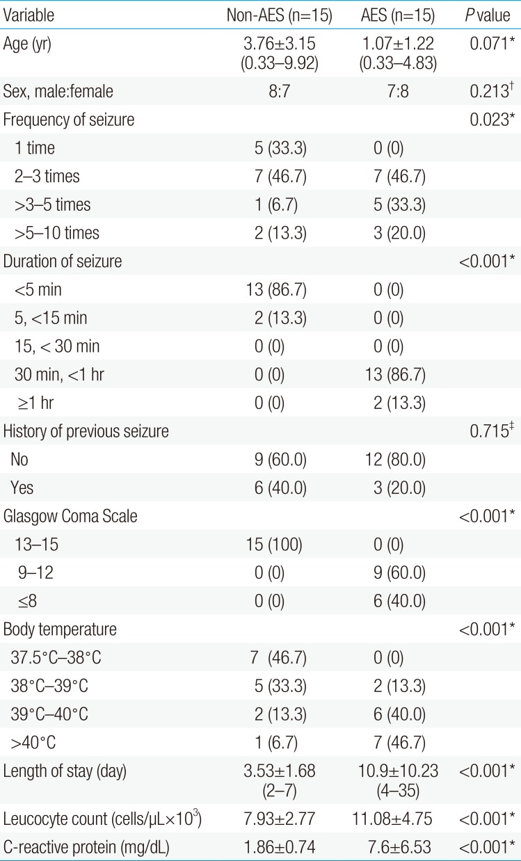
In the study period, 30 patients were enrolled. Glasgow Coma Scale mostly decreased in the AES group by about 40% in the level ≤8. All patients in the AES group suffered from status epilepticus and 46.67% of them had body temperature >40℃. Most of the cases in the AES group had longer duration of stay in the hospital. The median serum NSE level in the AES group was 157.86 ng/mL, and this value was significantly higher than that of the non-AES group (10.96 ng/mL; P<0.05).
Conclusion
AES cases showed higher levels of serum NSE. These results indicate that serum NSE is a good indicator of neuronal brain injury.
Introduction
Acute encephalitis syndrome (AES) is one of pediatric neurology health problem in Indonesia1). AES is a group of clinically comparable neurologic symptom caused by a number of different bacteria, viruses, fungus, spirochetes, parasites, chemical/toxins etc.23). There is seasonal and geographical variation in the causative organism. There were 165,461 cases from 1978 to 2011, of which and 40,431 were from Nepal and 125,030 cases from India. The mean incidence rate of India was 0.42 (standard deviation [SD], ±0.24) and that of Nepal was 5.23 (SD, ±3.03)4). AES is a constellation of clinical signs and/or symptoms, i.e., acute febrile disease, with an acute change in mental status and/or new onset of convulsions23). These clinical findings represent the patient has acute inflammation of the brain and are used by clinicians to identify patients with acute encephalitis. Acute encephalitis can be related with severe complications, including seizures, limb paresis, impaired consciousness or death4567). Biomarker of neuronal damage in intracranial infection is importance to recognize to predict outcome and disease severity.
Neuron specific enolase (NSE) is generally used as a marker for the neuropathological process in the brain. NSE is a glycoprotein enzyme present almost exclusively in neurons and neuroendocrine cells. It is a protein that is functionally active as heterodimer assembled from a combination of three subunits: α, β and γ8910). NSE is located in the cytoplasm of the neurons. The biologic half-life of NSE in the serum is 24 hours. It was first used as a tumor marker for small-cell lung cancer, neuroblastoma and other malignancies of neuroendocrine origin, and later it was introduced as a marker for brain damage. Due to obvious ethical obstacles for performing cerebrospinal fluid (CSF) collection in some neurological disorder, several studies have suggested the use of serum NSE to estimate neuronal damage91011). Hence, the interpretation of research using only serum NSE has been controversial. Some have provided positive results while others have not. Nevertheless, up to this date, the number of studies monitoring AES patients using serum biomarkers is limited.
The aim of the study is to investigate whether serum NSE was expressed in AES that cause neuronal damage in children.
Materials and methods
1. Patients
The regional ethics committee of Soetomo Hospital approved this prospective observational study. The study was conducted from July to December 2015. Comprehensive informed consent was obtained from the legal representative of the patient. A case of AES is described as a of any age, at any time of year with the acute onset of fever and alteration in mental status (including manifestation such as disorientation, confusion, inability to talk or coma) and/or new onset of convulsion (excluding simple febrile convulsion)23). Other early clinical information may include an increase in irritability, somnolence or unusual behavior greater than that observed with usual febrile illness23). Cases of AES at the age of 1 month to 12 years were included in the study. Cases that categorized as simple and complex febrile seizures were considered as non-AES group. The patients were excluded if they had history of intracranial tumors, head trauma, hydrocephalus and motoric neurological dysfunction.
2. Methods
In every patient, Glasgow Coma Scale (GCS) was assessed at admission in the Emergency Department before sedation was started. Within 24 hours after hemodynamic stabilization, 1 mL blood was drawn from a radial arterial catheter for the measurement of NSE. The outcome was determined upon discharge.
3. Measurement of NSE
All blood samples were immediately centrifuged and aliquoted at −70℃ until analysis was performed. NSE was measured with a Human NSE ELISA Kit (Elabscience Biotechnology Co., Ltd., Houston, TX, USA), by using spectrophotometers in 450-nm wavelength. The results were stated in ng/mL, with normal NSE value in children 7.2–12 ng/mL.
4. Statistical analysis
SPSS ver. 12.0 (SPSS Inc., Chicago, IL, USA) was used for statistical analysis. Chi-square test, Fischer exact test, and Mann-Whitney U test were applied to evaluate data comparison between groups. Mann-Whitney test was used for comparison of median NSE value between the 2 groups. Statistical significance was considered at 2-sided P value of <0.05.
Results
In the study period, 30 patients, 15 male and 15 female patients ranging in age from 3 months to 9 years were enrolled in the study. Two patients were excluded because of technical difficulty on blood taken samples and insufficient blood sample volume. There was no difference in age, sex and frequency of seizure between groups.
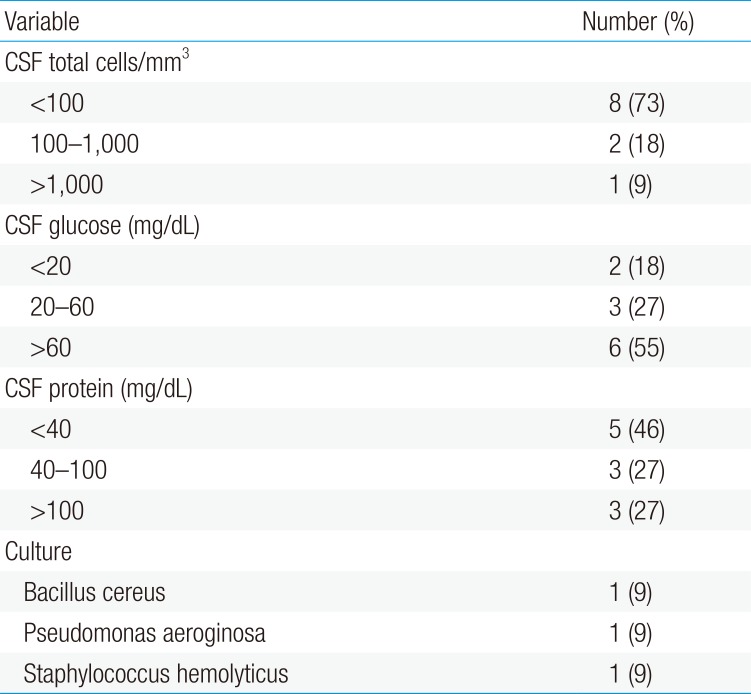
Consciousness that assessed with GCS mostly decreased in AES group for about 40% in the level ≤8. All patients in AES group suffered from status epilepticus and 46,67% of them with temperature >40℃. Most of AES group show longer duration length of stay in the hospital and higher level of CRP and leucocyte count as shown in Table 1. Most of CSF analyses showed viral infection with 3 types of bacteria were isolated from the CSF as shown in Table 2. Table 3 show a comparison between the median serum levels of NSE in children with AES and non-AES. The median serum NSE in AES group was 157,86 ng/mL, this value was significantly higher than serum NSE in non-AES group which was 10,96 ng/mL. There was significant correlation between serum NSE level and motor deficit outcome in AES as shown in Table 4.
Discussion
This study showed a remarkable change of serum NSE level in AES compared to non-AES. The ideal biomarker for brain injury will provide (1) specificity: it will be uniquely present in the central nervous system and accurately demonstrate the extent of brain injury, (2) sensitivity: it would be highly abundant and easily detectable, and (3) determine therapeutic effectiveness: biomarker levels should reflect successful therapeutic intervention. In this concept in mind, we will focus on structural protein biomarker measured from the blood8). Given ethical restrictions and limitations of lumbar puncture, estimation of serum NSE would be an available selection than CSF NSE. Literature documents have shown inconsistencies among investigation concerning the exclusive use of serum NSE to estimate neuronal damage. One could expect that serum NSE could replace CSF NSE by at least in cases which present blood-brain barrier (BBB) disruption9).
The existing findings indicate that patients in the AES group had higher serum NSE levels than other groups. The AES group was characterized by a severe neurological disorder that had in common lower scores on GCS. The disturbance of integrity of BBB may NSE reach the serum compartment. These results indicate that serum NSE provides a good indicator of brain injury, as suggested by the previous study. This study is contrary with a study by Lima that indicates that serum NSE is not sensitive enough to detect neuronal damage, but CSF NSE seems to be a consistent parameter for estimating patients with considerable neurological injury as well as cases with adverse outcome. Another restriction of the interpretation of serum NSE levels is the putative presence of hemolysis that could lead to false-positive results9). Bartek et al.12) report that cerebral metabolism is frequently affected in patients with acute bacterial meningitis. Furthermore, patient with higher lactate:pyruvate ratio had significantly higher levels of NSE, suggesting an ongoing deterioration in compromised cerebral tissue. As reported previously in the literature, interpretation of biomarkers may help the clinician to optimize the treatment strategy in cerebral pathologies, limiting the effects of inconsiderable insults and therefore theoretically prevent the progress of existing brain injury. In children with acute viral encephalitis following hand-foot-mouth diseases, there was significantly higher level of serum NSE compared to nonencephalitis group13). Brain neurotransmitter dysfunction is involved in septic encephalopathy. Increased BBB perimicrovascular edema associated with astrocyte injuries and invasive intracerebral infections were found in the first hours after inducing sepsis. Brain dysfunction was a component of multiple organ dysfunction syndrome, as reflected by the highest biomarker values14). NSE increased in a more variable way in sepsis due to its longer half-life, but persisting high levels could indicate brain inflammation and neuronal death14).
The potential for brain injury to occur following seizures is a topic of great interest and controversy. Although animal studies suggest that seizures in themselves lead directly to neuronal death, interpretation of human data is often complicated by numerous factor15). Our result showed all patients with AES suffered from status epilepticus. NSE is a marker of brain injury and BBB dysfunction, which is elevated following seizures in adults, but moderately few data exist on postictal NSE levels in children. NSE serum levels were substantially higher in children with status epilepticus (SE) and there was significant correlation between the seizure duration and the serum levels of NSE16). Elevated NSE serum levels could be demonstrated as a consequence of ischemia which results from prolonged seizures and induced cytoplasmic loss of NSE in the nervous system neurons, corresponded quantitatively to the severity of neuronal injury and duration of seizures, and were detectable before irreversible neuronal injury9). Experimental studies demonstrated changes in permeability of the BBB in animals experiencing long-lasting seizures. Human studies described in vivo that after SE, there is a marked dysfunction of the BBB as measured by QAlb (serum/CSF albumin ratio)17). The demonstration of an elevated QAlb in SE patients supports the notion that the increase of NSE is most probably regulated by the degree of permeability of the BBB17). In contrary, less than 10% of children had elevated NSE level following a seizure. Elevated NES level occurred only in children with severe neurologic disorders, in whom it was difficult to determine whether the seizure, the underlying etiologic process, or some other complicating factor was most critical in producing the neuronal damage15). There was limited study that correlates between nonspecific inflammatory markers with NSE in AES. Our study showed mild elevated of mean white blood cell count and C-reactive protein (CRP) in AES group compared to non-AES group. Increased CRP production is an early and sensitive response to most form of microbial infection and the value of its measurement in diagnosis and management of various infective conditions has been established18). Pandey et al.19) stated that serum NSE and CRP concentration were found significantly higher in acute strokes. Both biomarkers were found significantly correlated with neurological disability and short-term outcome.
Our study showed a significant correlation between serum NSE level and motor deficit outcome in AES. Comparing of the result with non infectious disease outcome, serum levels of NSE in first few days of ischemic stroke can serve as a useful marker to predict stroke severity and early functional outcome20). Study by Rech et al.21) demonstrates that NSE levels measured early in the course of ischemic cerebral injury are significantly higher in patients with unfavorable outcome than in patients with favorable outcome.
Several limitations of the recent study must be considered. Brain injury was only demonstrated in a limited number of patients due to practical and clinical safety conditions. Apparently, a single determination of serum NSE offers limited information. Ideally, to better understand the exact relation between the concentrations of NSE in the CSF compartment and the serum compartment, CSF and serum should be consecutively sampled. However, a serial collection of CSF samples in AES is rarely clinically indicated.
In conclusion, AES cases showed a higher value of serum NSE level. These results indicate that serum NSE provides a good indicator of neuronal brain injury. Although NSE should be expected to detect necrotic cell death, the sensitivity of NSE for assaying apoptotic cell death and nonlethal neuronal injury is uncertain. Further clinical studies of infection related neuronal injury in children, using NSE and other assays of neuronal damage, should help target the most appropriate populations for neuroprotective strategies.
Acknowledgments
We are thankful for Juhdy Husnudin for helping data collection.
Notes
Conflicts of interest: No potential conflict of interest relevant to this article was reported.


