Correlation of serum S100B levels with brain magnetic resonance imaging abnormalities in children with status epilepticus
Article information
Abstract
Purpose
To evaluate the association between elevated S100B levels with brain tissue damage seen in abnormalities of head magnetic resonance imaging (MRI; diffusion tensor imaging [DTI] sequence) in patients with status epilepticus (SE).
Methods
An analytical observational study was conducted in children hospitalized at Dr Soetomo Hospital, Surabaya, from July to December 2016. The patients were divided into 2 groups: SE included all children with a history of SE; control included all children with febrile seizure. Blood samples of patients were drawn within 24 hours after admission. SE patients also underwent cranial MRI with additional DTI sequencing. The Mann-Whitney test and Spearman test were used for statistical analysis.
Results
Fifty-three patients were enrolled the study. In the 24 children with SE who met the inclusion criteria, serum S100B and cranial MRI findings were assessed. Twenty-two children admitted with febrile seizures became the control group. Most patients were male (66.7%); the mean age was 35.8 months (standard deviation, 31.09). Mean S100B values of the SE group (3.430±0.141 μg/L) and the control group (2.998±0.572 μg/L) were significantly different (P<0.05). A significant difference was noted among each level of encephalopathy based on the cranial MRI results with serum S100B levels and the correlation was strongly positive with a coefficient value of 0.758 (P<0.001).
Conclusion
In SE patients, there is an increase of serum S100B levels within 24 hours after seizure, which has a strong positive correlation with brain damage seen in head MRI and DTI.
Introduction
Status epilepticus (SE) is neurological events causing significant morbidity and mortality in children [1]. Early definitions of SE described seizures persisting “for a sufficient length of time or is repeated frequently enough to produce a fixed or enduring epileptic condition [2]. In recent years, many studies have been conducted on biomarkers in biological fluids that correlate with neurological disorders, for example, neuron-specific enolase or Tau protein [3]. One of the most studied markers is the S100B protein [3-5]. The use of biomarkers mainly rests on the high sensitivity of markers that can be a clinician's reference in taking diagnostic and therapeutic action to patients.
Imaging examination in SE aims to evaluate structural damage occurred, which may indicate the cause of the seizure as well as the clinical prognosis of the patient. Magnetic resonance imaging (MRI) is very useful to know the existence of structural damage to the brain that occurred sometime after the incidence of hypoxia-ischemia [6]. The prognostic use of MRI is growing with diffusion tensor imaging (DTI) techniques that have enhanced the role of MRI in examining patients with global brain hypoxia-ischemia. MRI sequences with DTI sequences can detect central nervous system microstructures and detect undetectable abnormalities with conventional MRI findings [7].
The purpose of the research is to evaluate the association of elevated S100B levels with brain tissue damage seen in abnormalities of head MRI (DTI sequence) in patients with SE.
Materials and methods
1. Patients
We conducted an analytical observational study in children hospitalized at Dr Soetomo hospital, Surabaya from July 2016 to December 2016. The patients were divided into 2 groups. The SE groups consisted of all children with a history of SE that met the following criteria: (1) aged 1 month up to 18 years, (2) first experience of SE, (3) obtained parental consent indicated by filling out the written informed consent. SE characterized as any seizure lasting 30 minutes or more or intermittent seizures lasting for more than 30 minutes without recovery of consciousness interictally [2]. They were excluded if there were history of head trauma and congenital abnormalities. The control group consisted of all children with simple febrile seizure (FS). Simple FSs require all of the following features: a duration of less than 15 minutes, generalized in nature, a single occurrence in a 24-hour period, and no previous neurologic problems [8]. Epilepsy was defined as a chronic disorder of the brain characterised by an enduring disposition towards recurrent unprovoked seizures [9]. Meningoencephalitis was defined as acute infection of the brain in which the meninges, the subarachnoid space and the brain parenchyma are together involved in the inflammatory reaction [3].
2. Methods
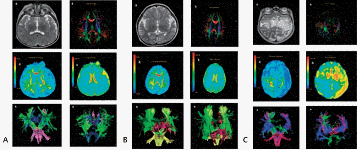
Blood samples of patients were drawn within 24 hours after admission, immediately sent for centrifugation, freezing (-80°C), and analyzed with Human S100B ELISA Kit (Elabscience Biotechnology, Houston, TX, USA), using a spectrophotometer with a wavelength of 450 nm. Patients with SE also underwent head MRI with an additional sequence of DTI taken with the MRT Optima 1.5T GE plane. Head MRI was performed 1 week following SE. DTI raw data processing performed by neuroradiology consultant, using GE advance windows workstation software (GE software volume share 5, GE Healthcare, Chicago Illinois, United States). MRI head abnormality is divided into 3 types as follows: grade I if the abnormality is limited to a part of white matter tract, grade II if there are lesions in the cortical and subcortical area, and grade III if there are lesions in most white matter (Fig. 1) [7]. All assessment results are recorded on the data collection sheets.
3. Ethical statement
The research ethical certificate was issued by the Health Research Ethics Committee, Dr. Soetomo Hospital, Surabaya with IRB 449/panke.KKE/VI/2016. The author received written informed consent from the patients.
4. Statistical analysis
IBM SPSS Statistics ver. 21.0 (IBM Co., Armonk, NY, USA) was used for statistical analysis. Mann-Whitney test was applied to compare S100B in both groups. Spearman test was used to correlate between variables. A 2-sided P value of <0.05 was considered statistical significance.
Results
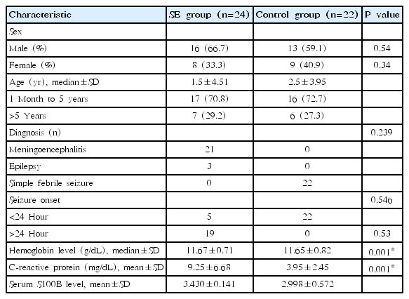
During research period, 53 eligible patients enrolled in the study. Four parents did not give consent and 3 patients died prior to head MRI. In 24 children who met the inclusion criteria, serum S100B and head MRI were assessed. Twenty-two children who admitted with FS became control group. We recorded demographic and clinical data in all subjects. Baseline characteristics of the subjects are shown in Table 1.
The S100B level in both groups did not have normal distribution. Mann-Whitney test was then used for statistical analysis. There was a significant difference between the S100B mean value of SE compared to the control group (Table 1). We evaluated MRI results and measured the fractional anisotropy and mean diffusivity parameters of patients in SE group.
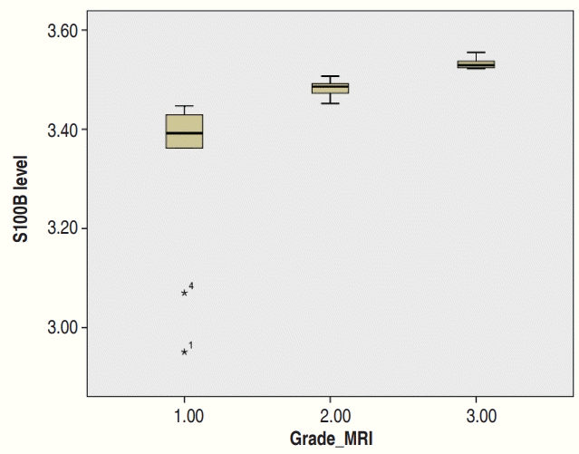
There is a significant difference in serum S100B levels between each level of encephalopathy based on head MRI results and the correlation is strongly positive (Table 2, Fig. 2) with coefficient value of 0.758 (P<0.001) with Spearman correlation test.

Analysis of S100B serum level in SE patients based on degree of encephalopathy noted on brain magnetic resonance imaging
Discussion
Elevated level of S100B was usually correlated with the underlying cause of seizure itself, such as subarachnoid hemorrhage, head injury, cardiac arrest and stroke [10-14]. In pediatric patients, S100B often investigated under conditions such as asphyxia, FSs, and intracranial infections [3,15-17]. This study shows elevated serum S100B levels significantly in SE group. S100B protein naturally present in the central nervous system due to the apoptosis of nerve cells. In the condition of the open blood barrier due to a central nervous system inflammation, whether caused by infection, hypoxic injury from seizures, or trauma, S100B protein will diffuse into the bloodstream and the level will increase in blood [18]. This characteristic makes the S100B an ideal marker to detect brain cell damage that occurs during SE.
Head MRI has been widely used as a means to establish diagnosis and etiology on the SE. MRI with DTI sequences is able to show the possibility of structural and functional damage. A study by Xie in China, using the DTI sequence MRI tries to evaluate the functionally and structurally disrupted brain regions in each type of epilepsy that affects the patient's level of consciousness, namely partial complex seizures, generalized tonic-clonic seizures, and generalized tonic seizures secondary clonics [18]. DTI is a fairly recent technique that belongs to an advanced MRI which can assess the movement of water. DTI can detect microstructure of the central nervous system and detect undetectable abnormalities with conventional MRI as well as have an advantage in describing the microstructure of brain tissue during the period of growth and maturation of the brain. Most abnormalities on MRI are focal and unilateral, limited to cortical to subcortical areas. Abnormalities can originate in the cerebral neocortex or from the posterior and perirolandic areas or peripheral parts of the old encephaloclastic lesions [19]. Other structures that can be affected include the contralateral cerebellar hemispheres, thalamus, splenium from the corpus callosum, caudate nucleus and globus pallidus [20]. Cortical laminar necrosis occurs because of the susceptibility of the cortical layer to anoxia and ischemia. Besides neurons, glia cells and blood also get damaged and cause a condition known as pan-necrosis. Selective susceptibility of gray matter is caused by higher metabolic requirements and denser excitatory amino acid receptor concentrations where amino acids will be released after anoxic-ischemic events.
This study shows strong positive correlation between S100B levels and degree of encephalopathy shown in head MRI. Serum S100B levels can illustrate the possibility of the risk of epileptogenesis in brain cell damage seen in contrast MRI images of the head. In a study conducted by Cianfoni in Italy, SE can trigger reversible changes and abnormalities in MRI images. Reversible changes in MRI images will last for 15–150 days with the average change occurring within 62 days. Conditions of SE will produce incomplete reversibility in the form of residual gliosis and focal atrophy on abnormal MRI images. This will be useful in determining differential diagnoses and preventing misdiagnosis that will affect management options for patients [21].
Many research has been done to determine the relationship between serum S100B levels and imaging modalities. Based on a study conducted by Calcagnile in Sweden, in patients with mild brain injury due to trauma examination plasma S100B levels can provide sensitive results as a negative, it is helpful in saving the cost of diagnostic and patient care [22].
Linsenmaier et al. [23] then tried to examine the relationship between the presence of intracranial nontrauma lesions in patients with elevated plasma S100B levels, and the relationship between S100B levels and intracranial bleeding due to minimal head trauma seen in head CT and MRI scans. It resulted that plasma S100B levels can be used instead of head CT scan in detecting the presence or absence of intracranial bleeding due to head injury. In contrast, examination of plasma S100B levels has a low specificity for the presence of traumatic intracranial structural abnormalities because plasma S100B levels can increase if nontrauma intracranial lesions are found such as brain atrophy, microangiopati, chronic parenchymal brain defect. This has resulted in the use of MRI as a diagnostic tool for brain imaging that is superior to head computed tomography scan is still needed in an effort to assess brain damage functionally or structurally [23]. S100B examination is considered to be quite useful in saving maintenance costs, especially for expensive diagnostic imaging tests. In Dr. Soetomo Hospital, the implementation of MRI imaging requires high financing (±US dollars [USD] 380), while for S100B test costs less than USD 35.
This study substantiates the previous research evidence of elevated serum S100B levels in nerve cell damage caused by hypoxia when SE occurred. Increased serum S100B levels can be a reference in predicting how much nerve cell damage is occurring, which in turn will be a consideration in planning the imaging examination as well as the treatment plan to be administered. The limitations of this study include a less varied population in which research cases limited to SE caused by meningoencephalitis, epilepsy and FSs, so the results cannot be generalized to other disorders. In addition, the seizure etiology cannot be fully controlled, so elevated levels of S100B may also be a direct result of the underlying disease of the patient.
In conclusion, there is an increase in serum S100B levels within 24 hours after seizure in patients with SE, and this has a strong positive correlation with brain damage seen in head MRI and DTI.
Notes
No potential conflict of interest relevant to this article was reported.
Acknowledgements
We are thankful for all the staff Dr Soetomo hospital for helping data collection.


