Warning: fopen(/home/virtual/pediatrics/journal/upload/ip_log/ip_log_2024-12.txt) [function.fopen]: failed to open stream: Permission denied in /home/virtual/pediatrics/journal/ip_info/view_data.php on line 93

Warning: fwrite(): supplied argument is not a valid stream resource in /home/virtual/pediatrics/journal/ip_info/view_data.php on line 94
Strategies to support language development in neonatal intensive care unit: a narrative review

Strategies to support language development in neonatal intensive care unit: a narrative review

Article information

Clin Exp Pediatr. 2024;67(12):651-663
Publication date (electronic) : 2024 November 6
doi : https://doi.org/10.3345/cep.2024.00087
1Department of Pediatrics, Seoul National University College of Medicine, Seoul, Korea
2Department of Pediatrics, Seoul National University Children’s Hospital, Seoul, Korea
Corresponding author: Ee-Kyung Kim, MD, PhD. Department of Pediatrics, Seoul National University Children’s Hospital, 101 Daehak-ro, Jongno-gu, Seoul 03080, Korea Email: kimek@snu.ac.kr
Received 2024 January 12; Revised 2024 May 18; Accepted 2024 June 23.

Abstract

Despite neonatal intensive care advancements and quality improvements, preterm infants often experience delays in speech and language development during early childhood. The etiological pathway of language delays is multifactorial, including younger gestational age at birth, male sex, pregnancy complications including gestational diabetes mellitus and preeclampsia, organic pathology from neonatal morbidities, environmental factors of the neonatal intensive care unit (NICU) and prolonged hospitalization, home environment including socioeconomic status and parental education, and parent-infant interactions. As early language experiences and environments are crucial for the development of language processing, strategies to support language development should be implemented from the NICU onward. This study aimed to summarize evidence- based strategies for language development through an extensive review of nutrition, NICU environment, language and sound exposure, developmental care interventions, and family-centered care. Promoting breastfeeding, increasing parent-infant interactions in a single-family room setting, nurturing the language environment via parental book reading and language interventions, and parent-integrated interventions in the NICU could potentially enhance language development among preterm infants. These supportive strategies can be integrated through family-centered care, which recognizes parents as primary caregivers and collaborative partners.

Key message

· Preterm infants often experience speech and language development delays during early childhood, impacting children's ultimate outcomes.

· Promoting breastfeeding, increasing parent-infant interactions in a single-family room, promoting a nurturing language environment by parental book reading and language interventions, and parent-integrated interventions in the neonatal intensive care unit could potentially enhance children's language development.

· Integrating these strategies through family-centered care is essential.

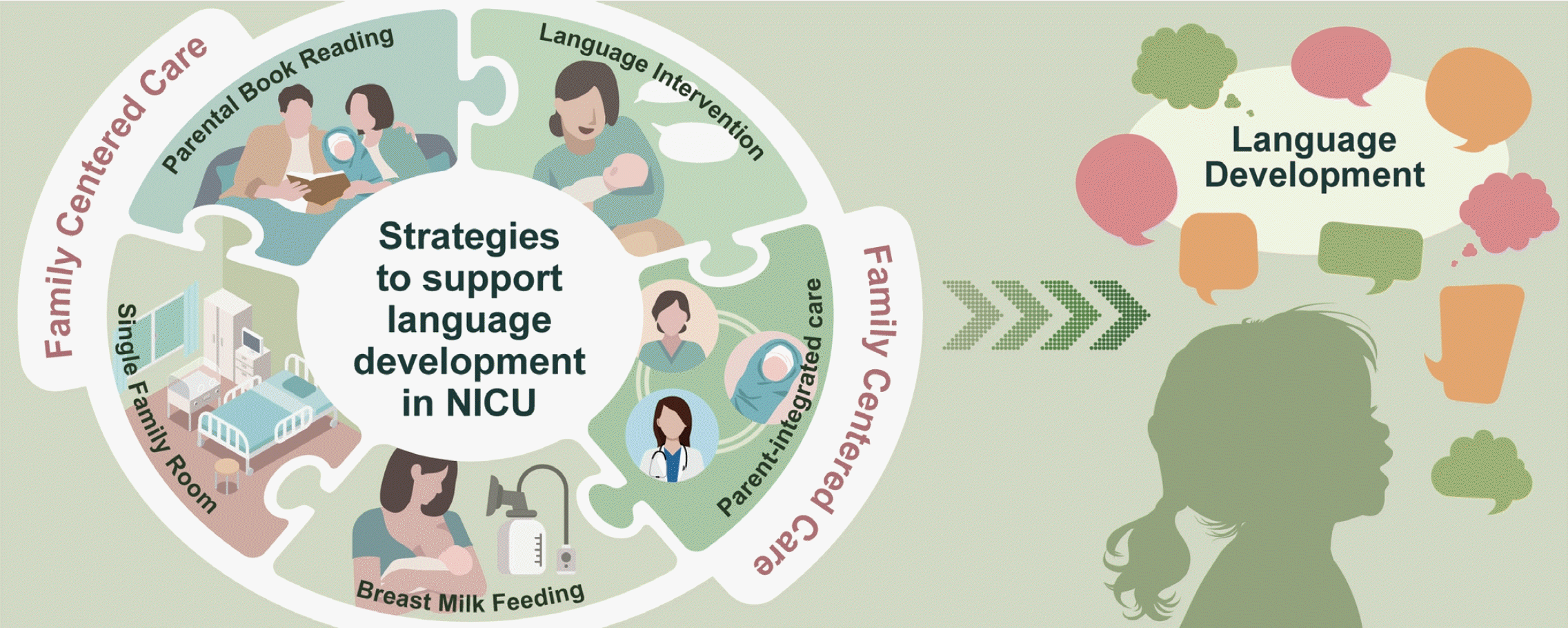
Graphical abstract. Strategies for supporting language development in neonatal intensive care unit (NICU).

Introduction

Early language development is crucial because it serves as the foundation for basic communication, cognitive process, literacy, and social interactions [1]. Language delays have profound and lasting impacts on children's ultimate outcomes. Preterm infants often face speech and language delays during early childhood that are often associated with other deficits including hearing loss, cognitive deficits, behavioral abnormalities, and cerebral palsy [2-4]. A prospective Canadian national cohort study demonstrated that language delays, defined as Bayley Scales of Infant and Toddler Development, 3rd edition (BSID-III) composite scores of <85, occurred in 35.3% of very preterm children [5]. According to a recent large-scale study of a national Korean cohort, the risk of developmental delay occurring by 6 years of age increased with younger gestational age at birth. In particular, preterm infants born before 28 weeks' gestation had a 3.57× higher risk of language disorders than full-term infants [6]. Since language relies on the brain's complex structure and function, alterations in which are significant indicators of neurodevelopment impairment [7]. Although survival and severe neurodevelopmental impairment have improved due to neonatal intensive care advancements and quality improvements, minimal advancements in language difficulties have been noted [8].

The etiological pathway of language delays is most likely multifactorial, including younger gestational age at birth and male sex [5,6,9]; pregnancy complications including gestational diabetes mellitus and preeclampsia [7]; organic pathologies from neonatal morbidities including bronchopulmonary dysplasia, brain injury, necrotizing enterocolitis and severe retinopathy of prematurity [5,9-12]; neonatal intensive care unit (NICU) environmental factors and prolonged hospitalization [13-15]; home environment including socioeconomic status and parental education [1,9,16-18]; multicultural families [19,20]; and parent-infant interactions [21-26]. Because early language experience is necessary for normal speech and language processing development, limited early language exposure in the NICU can be a significant cause of language delays among preterm infants [13,27,28].

Neuroplasticity, also known as brain plasticity, refers to the ability of the nervous system to change its activity in response to intrinsic or extrinsic stimuli by reorganizing its structure, functions, or connections [29]. Neuroplasticity is among the primary mechanisms through which humans adapt to environmental changes and demonstrate resilience to adverse events [4]. There may be a “sensitive period” in which neural circuits are particularly sensitive to environmental pressures [30]. In particular, the preterm and newborn periods, during which the development of brain connections and synaptogenesis occurs vigorously, involve greater plasticity [31]. The high plasticity of the brain during these early stages implies that positive experiences provided in an infant's environment can provide opportunities to positively influence cognitive and language outcomes [32]. For this reason, providing a nurturing language environment and appropriate access to aural or visual language in the NICU are critical for language development in preterm infants [21].

Broadly, this review centers on strategies that support language development in the NICU. Moreover, it provides an abridged overview of nutrition, language and sound exposure, parent-integrated interventions, and family-centered care in the NICU environment.

Nutrition

Early nutrition is critical to an individual's growth and development, and exclusive breast milk (BM) feeding is universally recommended for the health and well-being of all infants [33]. Preterm infants are at high risk for caloric and nutrient deficiencies, which can negatively affect their development. The critical nutrients found in BM, such as polyunsaturated fatty acids (PUFAs) and other neurotrophic factors, could have positive impacts on the brain development of preterm infants. Therefore, BM for preterm infants is emphasized even more.

The impact of BM, donor BM, and supplemental docosahexaenoic acid (DHA) provided to either mothers or neonates on infants' language development will be reviewed below.

1. Breast milk

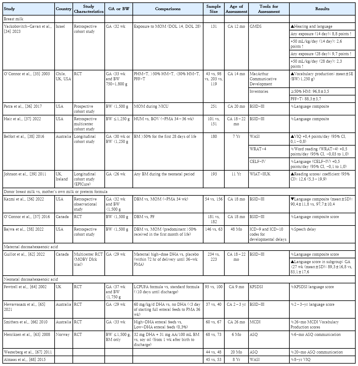
Several studies have assessed language outcomes following BM exposure in the NICU. Table 1 presents the impact of BM based on assessment age. Studies investigating the impact of BM on the language development at 12–14 months of corrected age (CA) reported positive effects [34,35]. In particular, Yackobovitch-Gavan et al. [34] reported that early exposure to the mother's own milk (MOM) at postnatal days 14 and 28 enhances hearing-language scores at a CA of 12 months in very preterm infants. Studies evaluating language developmental outcomes at a CA of 18–22 months using the BSID-III found no significant differences in language composite scores between the BM and non-BM groups [36,37]. Belfort et al. [38] evaluated the associations of BM intake by very preterm infants with neurological outcomes at 7 years of age. They concluded that predominant BM feeding in the first 28 days of life was associated with a better verbal intelligence quotient. However, no significant correlation was noted between breastfeeding and reading or language scores. In Johnson's study of the early predictors of educational outcomes of extremely preterm infants at 11 years of age, the receipt of any BM during the neonatal period was an independent predictor of high reading scores [39].

Studies evaluating language outcomes by NICU-provided nutrition

Limitations were noted in assessing the impact of breastfeeding during the early postnatal period due to inter-study variations among study subjects; breastfeeding definitions, timing, and duration; neurodevelopmental evaluation timing; and assessment tools. Nevertheless, the effect of BM seems stable from early life throughout childhood.

While the mechanisms explaining the relationship between BM and language development have not been extensively researched, several possibilities should be considered. Postnatal brain development may be sensitive to dietary elements because of the relatively permeable nature of the infant gut and blood-brain barrier to small molecules [40]. Previous studies demonstrated that BM affects neural tissue structure and gray- and white-matter maturation and myelination, leading to favorable neurocognitive outcomes [38,41-44]. The fatty acid profiles of BM might play a significant role in brain structure and function. PUFAs, especially omega-3 (DHA) and omega-6 (arachidonic acid), are involved in neurogenesis, neuronal migration, gliogenesis, synaptogenesis, and myelination from fertilization [45-48]. This process may shape the neural pathways linked to the developmental progression of language acquisition [49].

Other constituents of BM, including micronutrients and bioactive components such as human milk oligosaccharides, hormones, growth factors, and immunoglobulins, may contribute to brain development and work synergistically to optimize neurodevelopmental outcomes [50-53]. Moreover, BM may impact neurodevelopment via epigenetic mechanisms. Its bioactive compounds, such as noncoding RNAs, stem cells, and microbiomes, are under study as epigenetic modulators that influence neurodevelopment [54]. Furthermore, breastfeeding, whether directly or via exclusive pumping, stimulates the secretion of maternal pituitary hormones, particularly oxytocin. These oxytocin changes persist for an extended period and may affect maternal behavior and the motherhood experience as well as influence infant cognition, learning, and language developmental outcomes [51,55].

2. Donor BM

With an increasing awareness of the positive effects of BM on neonatal growth and development, the use of pasteurized donor BM has become increasingly popular in the NICU when the MOM is unavailable. Several studies have assessed the long-term effects of donor BM on language development (Table 1). Kazmi et al. [56] reported that infants fed donor BM had lower language scores at CA of 18 months compared to those fed MOM. In a randomized controlled trial (RCT) comparing donor BM and preterm formula for 90 days or until discharge, the use of supplemental donor BM did not improve language development among very low birth weight (VLBW) infants at a CA of 18 months [57]. In a retrospective cohort study evaluating neurodevelopment at 48 months of age, no significant differences were found in the risk of speech delay between infants who received donor BM versus MOM [58]. Thus, donor BM did not have a positive impact on long-term language development.

Donor BM may not offer similar developmental benefits to MOM due to several factors, including the compositional differences between the milk of mothers of preterm versus full-term infants [59,60], the loss of certain nutrients during the pasteurization, storage, and processing of donor BM [59,61], and the lack of hormonal changes in the mother with milk expression.

3. Docosahexaenoic acid

Lipids, accounting for more than 50% of the cerebral tissue’s dry weight, are the nutrient deemed most closely related to brain development. DHA (C22:6n-3) is an essential long-chain PUFA in nervous system formation and function, particularly the brain and retina. DHA is currently regarded a crucial nutrient during pregnancy and breastfeeding due to its active involvement in early nervous system development [47].

Maternal DHA supplementation

A multicenter RCT evaluated the effect of maternal high-dose DHA on the language neurodevelopment of very preterm infants at a CA of 18–22 months (Table 1) [62]. Maternal DHA supplementation did not improve the language outcomes of breastfed preterm infants. However, a subgroup analysis suggested a potential benefit of maternal DHA for the language development of preterm infants born before 27 weeks’ gestation. Additional well-designed research is needed to confirm this finding.

Neonatal DHA supplementation

Table 1 shows several RCTs that evaluated the effects of neonatal DHA supplementation on language developmental outcomes. Two demonstrated neurodevelopmental effects within 1 year of age [63,64]. Henriksen et al. [63] reported that DHA supplementation for BM-fed preterm infants did not improve the communication subtest scores of the Ages and Stages Questionnaire. Fewtrell et al. [64] also showed no significant difference in Knobloch, Passamanick and Sherrard’s Developmental Screening Inventory language scores between long-chain PUFA and standard formula-fed groups. In studies assessing language development at a CA of 18 months to 3 years, no significant differences in developmental scores were noted between the groups supplemented withDHAand the control group [65-67]. Almaas et al. [68] administered the Wechsler Abbreviated Scale of Intelligence to 8-year-olds to evaluate the long-term effects of DHA supplementation during NICU hospitalization. Their findings revealed no significant difference in verbal intelligence quotient between the DHA group and the control group. Summarizing the current research findings, it is challenging to consider neonatal DHA supplementation as having positive effects on the language development of preterm infants.

NICU environment

The structural components of the ears develop in the first 20 weeks of gestation [69]. The human cochlea and peripheral sensory end organs normally develop fully by 24 weeks' gestation [70]. The auditory system typically becomes functional around 25 weeks' gestation. The hearing threshold is approximately 40 decibels A (dBA) at 27–29 weeks' gestation. It gradually decreases to a nearly adult level (13.5 dBA) by 42 weeks' gestation, indicating ongoing postnatal maturation of the auditory pathways of the central nervous system [71]. The period from 25 weeks' gestation to 5–6 months of age is crucial to the development of the neurosensory part of the auditory system [69].

The language and sound environment to which fetuses and preterm infants are exposed differs. The intrauterine environment is characterized by constant primarily low-frequency sounds from the mother's cardiovascular and digestive systems transmitted through the amniotic fluid [72,73]. Extrauterine sounds are altered as they pass through abdominal tissue, leading to some sound attenuation that may be more pronounced at high- and mid-frequency energy bands [72,73]. Language and extrauterine sound exposure for fetuses exhibited a marked cyclical day/night pattern with reduced exposure during nighttime hours [74]. In contrast, preterm infants in the NICU are minimally exposed to biological sounds; in fact, they may receive nearly 5×s less language exposure than fetuses [74]. Instead, they are exposed to high sound levels and electronic and mechanical noises, all of which are transmitted through the air rather than through fluid [75,76]. Such exposures vary significantly less throughout the 24-hour cycle [74]. These abnormal auditory exposures for preterm infants may contribute to speech/language deficits later in life [13,77].

1. Single-family room versus open-bay NICU

The traditional NICU was an open-bay environment of multiple beds located in a single space. However, most human conversations in an open-bay NICU design are blocked by ambient noise, which deprives infants of meaningful language stimulation. As a result, recent recommendations for NICU design include the construction of private spaces, referred to as single-family rooms (SFRs), to reduce ambient noise and facilitate family involvement [78]. However, single-room NICU designs also feature increased social isolation, causing language deprivation and atypical language development [13]. Therefore, it is important to evaluate language outcomes comparatively by NICU design.

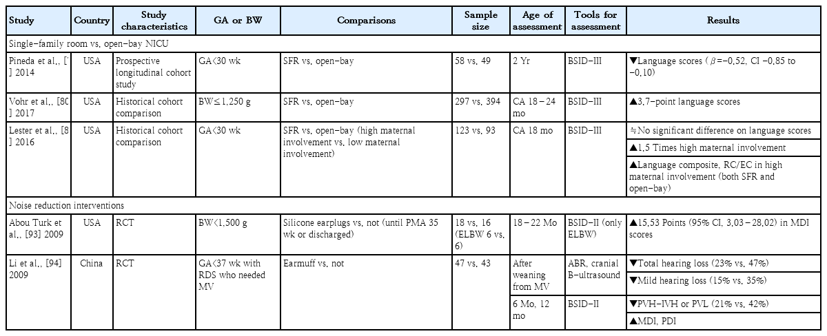
Regarding long-term language outcomes, several studies reported conflicting results (Table 2). In the prospective longitudinal cohort study of Pineda et al. [79], very preterm infants cared for in SFRs had reduced cerebral maturation at term-equivalent age and poorer language development at 2 years compared to those cared for in open bays. On the other hand, Vohr et al. [80] reported that the SFR NICU was associated with a 3.7-point increase in mean language score at a CA of 18–24 months. Lester et al. [81] reported that infants with high maternal involvement in the NICU (both SFR and open-bay designs) had greater language composite, receptive, and expressive communication scores than infants with low maternal involvement. However, the number of days of maternal involvement and proportion of high maternal involvement were greater in the SFR than open-bay NICU design. The current evidence is insufficient to support any specific NICU design due to lack of RCTs. Nevertheless, due to the significant positive impact of SFR on language development, driven by increased maternal involvement through family-integrated care, it is reasonable to actively consider SFR design for future NICU facilities [81,82].

Studies evaluating language outcomes by NICU environment

2. Noise-reduction interventions

Overstimulation by the loud and noxious noises of the NICU has been well documented. According to American Academy of Pediatrics recommendations, safe sound levels in the NICU should not exceed an hourly level of 45 dBA [70]. However, the NICU noise level typically exceeds the recommended standards, often falling outside the acceptable range more than 70% of the time [75,83]. Long-term average equivalent sound levels (hourly, daily, or weekly) typically range from ~50 to 65 dBA in various NICU settings [75,84,85]. Short-term-equivalent sound level measurements can peak at 75–85 dBA [70,86]. This loud and noxious noise not only has negative short-term effects on the infant's cardiovascular and respiratory system, but it also exerts negative long-term effects on brain maturation and neurodevelopment [87-89]. Therefore, efforts to reduce noise in the NICU continue [90-92].

Only 2 studies have examined the long-term effects of noise reduction on the neurodevelopment of preterm infants (Table 2) [93,94]. One high-quality RCT that used silicone earplugs to reduce the sound level for VLBW infants showed the positive effects of earplugs on the Mental Developmental Index (MDI) at 18–22 months, especially only for extremely low birth weight infants [93]. However, the sample size was very small, with only 6 infants each in the earplug and control groups. In another RCT of preterm infants diagnosed with respiratory distress syndrome requiring mechanical ventilation, the group using earmuffs showed a significantly lower incidence of hearing loss and periventricular/intraventricular hemorrhage or periventricular leukomalacia than the control group. Additionally, the MDI scores at 6 and 12 months were significantly higher in the earmuffs group [94]. However, due to the small sample size, heterogeneous subjects, and variety of study methods used, it is difficult to draw a definitive conclusion regarding the noise-reduction effects of earplug or earmuff usage on language outcomes for preterm infants. Larger and well-designed trials are needed to verify results to date.

Language and sound exposure

During the last trimester of pregnancy, hearing becomes functional as previously discussed. In utero, low-frequency components of language, including pitch, as well as some aspects of prosody and rhythm are transmitted to fetuses through the uterus. Moreover, fetuses are exposed to properties of native language from their mother’s speech via bone conduction, aiding in early language acquisition [95]. This prenatal language experience shapes the neonatal neural response to speech [96]. Shortly after birth, neonates demonstrate specific responses to speech and can discern different prosodic patterns, indicating early tuning to the language environment [96,97]. Neuroimaging studies have provided considerable data about language development in neonates and young children. Perani et al. [98] demonstrated that, in 2-day-old infants, the language-related neural substrate was fully active in both hemispheres with a preponderance in the right auditory cortex. Neuroimaging conducted of 3-month-old infants indicated that speech processing relied on inferior frontal and temporal brain regions interconnected by 2 primary fiber bundles: the arcuate fasciculus and the uncinate fasciculus [99-101]. Minagawa-kawai et al. [102] delineated a clear left-lateralized cerebral basis for speech processing, predominantly to native language, in 4-month-old infants. Development of the speech production ability is associated with a notable increase in overall brain size. The most significant growth in cortical surface area occurs during the first and second years of life, with most notable growth of the superior temporal gyrus occurring during the first year of life [103,104]. In conclusion, a functional organization of the brain for language development is present from the fetal period to early infancy.

1. Parental book reading

Shared book reading is one of the most effective ways to enrich an infant's language and a well-recognized facilitator for higher language skills and literacy acquisition in preschoolers [105,106]. The American Academy of Pediatrics has issued a policy statement endorsing the commencement of parent-child home reading [107]. However, the impact of initiating parental book reading from the NICU setting on the subsequent language development of preterm infants with restricted language exposure remains unknown.

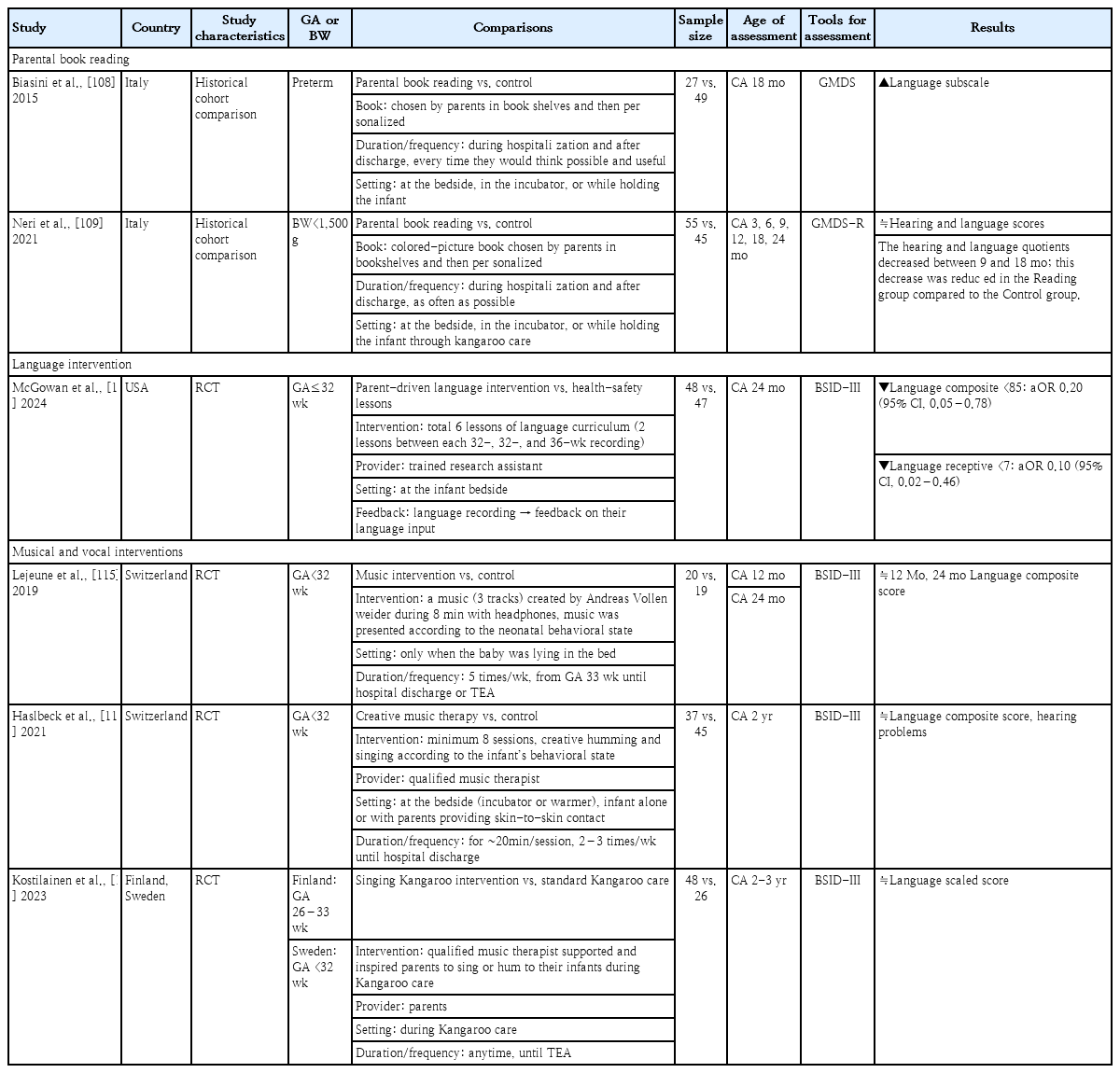
Two pre-post historical comparison studies from same institution evaluated the effects of parental book reading on the language development of preterm infants (Table 3) [108,109]. In both studies, the books were chosen by parents from bookshelves and then personalized. Parental book reading was conducted during the hospitalization, and the parents were encouraged to continue the practice after discharge. The frequency of book reading was not predetermined by a protocol; instead, the parents read books as often as possible. This was done at the bedside, in the incubator, or while holding the infant through kangaroo care. Biasini et al. [108] reported improved language scores for preterm infants at a CA of 18 months in the parental book reading versus control group. Neri et al. [109] examined VLBW infants and found no significant intergroup differences in hearing and language scores until a CA of 24 months. Both groups showed decreasing hearing and language quotients at a CA of 9–18 months. However, this decrease was reduced in the parental reading versus control group.

Studies evaluating language outcomes by language and sound exposure

Parental book reading has positive effects on language development, not only by exposing infants to their parents' language but also by positively influencing the parent-infant interaction, reducing parental stress, and enhancing functional brain connectivity [110-113]. Moreover, regular parental book reading may be an epigenetic factor associated with later reading success [107].

2. Language intervention

In a recent RCT, the language development of a parent-driven language intervention was compared with those of health-safety lessons (control group) (Table 3) [114]. The intervention group received 6 lessons of known methods for early language intervention involving language activities for parents to complete with their infants that included topics such as reading, singing, and narration of bedside activities. Natural language data recordings were obtained using a Language Environment Analysis digital language processor at 32, 34, and 36 weeks of life. This device analyzes the number of words spoken by adults, the child's vocalizations, and conversational turns. Parents in the intervention group received feedback on their language input to their infant every 2 weeks based on each previous language recording. The intervention group had higher adult word counts at 36 weeks and demonstrated an increase in adult word counts between recordings. The intervention reduced the risk of having a language composite score of <85 and a receptive score of <7 on the BSID-III assessment at a CA of 24 months. Thus, a language intervention in the NICU that leads to an increased exposure to adult word counts, may positively impact the language development of preterm infants.

3. Musical and vocal interventions

Three RCTs assessed neurodevelopment by applying music therapy or singing (Table 3) [115-117]. The enrolled study participants were younger than 32–33 weeks' gestation. One study provided recorded music [115], while the other 2 studies offered songs/humming provided directly by music therapists or the parents [116,117]. The interventional duration and frequency varied among the studies. When evaluating language development at a CA of 12 months and 2–3 years, the studies found no significant intergroup differences in language scores. This comprehensive review of current research findings identified no clear evidence to substantiate the effectiveness of musical and vocal interventions for preterm infants.

Parent-integrated interventions

The NICU environment can induce stress for both parents and infants through early separation, potentially impeding early bonding and attachment [118]. Considering that parents are invaluable as the primary caregivers of and crucial advocates for their infants, there is a strong emphasis on early involvement of parents in the NICU to support their engagement in early caregiving and promote an adaptive post-discharge transition to home [119-121].

Two RCTs evaluated the neurodevelopmental outcomes of early parent-integrated interventions in the NICU (Table 4) [122,123]. The Family Nurture Intervention was designed to overcome the maladaptive conditioning effects of maternal separation and the NICU environment on preterm infants [122]. Nurture specialists facilitated the Family Nurture Intervention during mother-infant sessions. The first activity was a scent cloth exchange between the mother and the infant. Nurture specialists encouraged the mothers to engage in touching, communication, and maximizing eye contact with their infants during both skin-to-skin and non-skin-to-skin holding sessions. The Family Nurture Intervention significantly improved the cognitive and language scores of infants whose scores were greater than 85 at a CA of 18 months [122].

Studies evaluating language outcomes by parent-integrated intervention

Another early parenting intervention consisted of 2 psychoeducational group sessions and 4 individual sessions [123]. The group sessions provided information about the typical characteristics of preterm infants, their special caregiving needs, and the procedures conducted by NICU staff. This information was delivered by a pediatric neuropsychiatrist and a neurodevelopmental therapist. The individual sessions consisted of the joint observation of the infant and communication skills to support parenting efforts. When assessing psychomotor development using the Griffith Mental Development Scales at a CA of 24 months, the intervention group demonstrated significantly higher global scores and hearing-speech scores than the control group [123].

Family-centered care

Considering the previously discussed content, it becomes evident that promoting breastfeeding, increasing parent-infant interactions in an SFR setting, encouraging parental book reading and language interventions, and facilitating parent-integrated interventions in the NICU may enhance the language development of preterm infants. All of these strategies can be implemented through family-centered care.

Family-centered care enables parents to act as the primary caregivers of their babies and partners with clinical teams. Key concepts of family-centered care include an environment that is designed to support 24-hour parental presence/participation, NICU team education and support, parent education/psychological support, and active parent participation/partnership [124,125]. There is growing evidence of the potential advantages of family-centered care of hospitalized preterm infants. Additionally, it enhances breastfeeding rates and contributes to increased weight gain [126-128]. Moreover, it reduces morbidities such as retinopathy of prematurity, bronchopulmonary dysplasia, infections, and intraventricular hemorrhage, leading to a decreased length of hospital stay. 126,127)Ultimately, family-centered care reduces parental stress and anxiety and improves neurodevelopmental outcomes, including language development [126,127,129,130].

Despite its benefits, implementing family-centered care in the traditional NICU is challenging due to difficulty changing the perceptions of healthcare professionals and parents, a shortage of well-trained specialists, and issues related to sustained fidelity. As a result, family-centered care is still not implemented in most NICU facilities in Korea. To implement family-centered care, a fundamental shift in mindset is required that moves away from professional-centered care and hierarchical hospital culture. Parents must be considered the primary caregivers and recognized as collaborative partners in all aspects of NICU care. There is also a need to develop early assessments and systematically tailored intervention programs to implement family-centered care plans. Adopting a multidisciplinary team approach, including speech therapists, otolaryngologists, neurologists, and rehabilitation medicine specialists, is crucial. This approach should also encompass parental education regarding language development and intervention along with programs aimed at providing emotional support to parents. Ultimately, it is crucial to develop standardized protocols and national policies, establish standardized assessment metrics, and ensure national financial support for the successful integration of family-centered care into the domestic health care system.

Conclusions

Preterm infants often face speech and language developmental delays of multifactorial etiologies. Promoting breastfeeding, increasing parent-infant interactions in an SFR setting, encouraging a nurturing language environment that consists of parental book reading and language interventions, and facilitating parent-integrated interventions in the NICU may enhance the language development of preterm infants. These supportive strategies can be integrated through family-centered care, which recognizes parents as primary caregivers and collaborative partners. These strategies for language development implemented in the NICU are expected to encourage a positive parent-child relationship and sustained language development efforts even after discharge.

Notes

Conflicts of interest

No potential conflict of interest relevant to this article was reported.

Funding

This study received no specific grant from any funding agency in the public, commercial, or not-for-profit sectors.

Author contribution

Conceptualization: JSH, EKK; Data curation: JSH; Formal analysis: JSH; Methodology: JSH, EKK; Project administration: JSH, EKK; Visualization: JSH; Writing-original draft: JSH; Writing- review & editing: JSH, EKK

References

1. McGowan EC, Vohr BR. Neurodevelopmental follow-up of preterm infants: what is new? Pediatr Clin North Am 2019;66:509–23.
2. Vohr B. Speech and language outcomes of very preterm infants. Semin Fetal Neonatal Med 2014;19:78–83.
3. Peralta-Carcelen M, Bailey K, Rector R, Gantz M. Behavioral and socioemotional competence problems of extremely low birth weight children. J Perinatol 2013;33:887–92.
4. DeMaster D, Bick J, Johnson U, Montroy JJ, Landry S, Duncan AF. Nurturing the preterm infant brain: leveraging neuroplasticity to improve neurobehavioral outcomes. Pediatr Res 2019;85:166–75.
5. Synnes A, Luu TM, Moddemann D, Church P, Lee D, Vincer M, et al. Determinants of developmental outcomes in a very preterm Canadian cohort. Arch Dis Child Fetal Neonatal Ed 2017;102:F235–4.
6. Jin JH, Lee SH, Youk TM, Yoon SW. Long-term outcomes of preterm infants in the first 6 years of life: a nationwide population-based study in Korea. Eur J Pediatr 2023;182:641–50.
7. Torres Y, Celis C, Acurio J, Escudero C. Language impairment in children of mothers with gestational diabetes, preeclampsia, and preterm delivery: current hypothesis and potential underlying mechanisms: language impartment and pregnancy complications. Adv Exp Med Biol 2023;1428:245–67.
8. Ricci MF, Shah PS, Moddemann D, Alvaro R, Ng E, Lee SK, et al. Neurodevelopmental outcomes of infants at <29 weeks of gestation born in Canada between 2009 and 2016. J Pediatr 2022;247:60–6.e1. .
9. Tseng WL, Chen CH, Chang JH, Peng CC, Jim WT, Lin CY, et al. Risk factors of language delay at two years of corrected age among very-low-birth-weight preterm infants: a population-based study. Children (Basel) 2023;10:189.
10. Schmidt B, Asztalos EV, Roberts RS, Robertson CM, Sauve RS, Whitfield MF. Impact of bronchopulmonary dysplasia, brain injury, and severe retinopathy on the outcome of extremely low-birth-weight infants at 18 months: results from the trial of indomethacin prophylaxis in preterms. JAMA 2003;289:1124–9.
11. Clark CA, Woodward LJ. Neonatal cerebral abnormalities and later verbal and visuospatial working memory abilities of children born very preterm. Dev Neuropsychol 2010;35:622–42.
12. Ahn JH, Lee KM, Kim MJ, Park HK, Kim YJ, Ahn SJ, et al. Neurodevelopmental outcomes in very low birthweight infants with retinopathy of prematurity in a nationwide cohort study. Sci Rep 2022;12:5053.
13. Rand K, Lahav A. Impact of the NICU environment on language deprivation in preterm infants. Acta Paediatr 2014;103:243–8.
14. Best K, Bogossian F, New K. Language exposure of preterm infants in the neonatal unit: a systematic review. Neonatology 2018;114:261–76.
15. Marston L, Peacock JL, Calvert SA, Greenough A, Marlow N. Factors affecting vocabulary acquisition at age 2 in children born between 23 and 28 weeks' gestation. Dev Med Child Neurol 2007;49:591–6.
16. Suskind DL, Leffel KR, Graf E, Hernandez MW, Gunderson EA, Sapolich SG, et al. A parent-directed language intervention for children of low socioeconomic status: a randomized controlled pilot study. J Child Lang 2016;43:366–406.
17. Valois R, Tojal C, Barros H, Costa R. [Formula: see text] Perinatal and social risk of poor language, memory, and learning outcomes in a cohort of extremely and very preterm children. Child Neuropsychol 2023;29:906–21.
18. Pan C, Gong Y. Association among postpartum posttraumatic stress disorder, family coping, neurodevelopment, and language development in high-risk infants: a retrospective study. Transl Pediatr 2022;11:728–37.
19. Chang YE, Sung MY, Chang MS, Kim SS, Lim AR. Analysis of trends and contents of multicultural early childhood language-related research. Multicult Peace 2019;13:40–63.
20. Kim HS, Kim HJ, Lee JW. A study on the trend of domestic research on the meta-language of children from multicultural families. Contemp Soc 2020;10:127–46.
21. Vohr BR. Language and hearing outcomes of preterm infants. Semin Perinatol 2016;40:510–9.
22. Nguyen TN, Spencer-Smith M, Haebich KM, Burnett A, Scratch SE, Cheong JLY, et al. Language trajectories of children born very preterm and full term from early to late childhood. J Pediatr 2018;202:86–91.e1.
23. Magill-Evans J, Harrison MJ, Van der Zalm J, Holdgrafer G. Cognitive and language development of healthy preterm infants at 10 years of age. Phys Occup Ther Pediatr 2002;22:41–56.
24. Sanchez K, Spittle AJ, Cheong JL, Thompson DK, Doyle LW, Anderson PJ, et al. Language in 2-year-old children born preterm and term: a cohort study. Arch Dis Child 2019;104:647–52.
25. Harel-Gadassi A, Friedlander E, Yaari M, Bar-Oz B, Eventov-Friedman S, Mankuta D, et al. Do developmental and temperamental characteristics mediate the association between preterm birth and the quality of mother-child interaction? Infant Behav Dev 2020;58:101421.
26. Ståhlberg-Forsén E, Latva R, Aija A, Lehtonen L, Stolt S. Language environment and parent-infant close contact in neonatal care and emerging lexical abilities of very preterm children-a longitudinal study. Acta Paediatr 2023;112:659–66.
27. Hersey A, Hoffman L, Tucker R, Vohr B. Enhancing the NICU language environment with a neonatal Cuddler program. J Perinatol 2021;41:2063–71.
28. Mayberry RI, Lock E, Kazmi H. Linguistic ability and early language exposure. Nature 2002;417:38.
29. Mateos-Aparicio P, Rodríguez-Moreno A. The impact of studying brain plasticity. Front Cell Neurosci 2019;13:66.
30. Tottenham N. The importance of early experiences for neuroaffective development. Curr Top Behav Neurosci 2014;16:109–29.
31. Kostovi I, Jovanov-Milosevi N. The development of cerebral connections during the first 20-45 weeks' gestation. Semin Fetal Neonatal Med 2006;11:415–22.
32. Spittle A, Orton J, Anderson PJ, Boyd R, Doyle LW. Early developmental intervention programmes provided post hospital discharge to prevent motor and cognitive impairment in preterm infants. Cochrane Database Syst Rev 2015;2015:CD005495.
33. World Health Organization. Infant and young child feeding. Geneva (Switzerland): World Health Organization; 2020. [cited 2023 Dec 13]. Available from: https://www.who.int/news-room/fact-sheets/detail/infant-and-young-child-feeding.
34. Yackobovitch-Gavan M, Atia Shmueli S, Morag I. Neurodevelopmental outcomes among infants born preterm fed with mother's own milk: a comparison of singletons and twins. J Pediatr 2023;259:113484.
35. O'Connor DL, Jacobs J, Hall R, Adamkin D, Auestad N, Castillo M, et al. Growth and development of premature infants fed predominantly human milk, predominantly premature infant formula, or a combination of human milk and premature formula. J Pediatr Gastroenterol Nutr 2003;37:437–46.
36. Patra K, Hamilton M, Johnson TJ, Greene M, Dabrowski E, Meier PP, et al. NICU human milk dose and 20-month neurodevelopmental outcome in very low birth weight infants. Neonatology 2017;112:330–6.
37. Hair AB, Patel AL, Kiechl-Kohlendorfer U, Kim JH, Schanler RJ, Hawthorne KM, et al. Neurodevelopmental outcomes of extremely preterm infants fed an exclusive human milk-based diet versus a mixed human milk+bovine milk-based diet: a multi-center study. J Perinatol 2022;42:1485–8.
38. Belfort MB, Anderson PJ, Nowak VA, Lee KJ, Molesworth C, Thompson DK, et al. Breast milk feeding, brain development, and neurocognitive outcomes: a 7-year longitudinal study in infants born at less than 30 weeks' gestation. J Pediatr 2016;177:133–9.e1. .
39. Johnson S, Wolke D, Hennessy E, Marlow N. Educational outcomes in extremely preterm children: neuropsychological correlates and predictors of attainment. Dev Neuropsychol 2011;36:74–95.
40. Engelhardt B. Development of the blood-brain barrier. Cell Tissue Res 2003;314:119–29.
41. Sullivan G, Vaher K, Blesa M, Galdi P, Stoye DQ, Quigley AJ, et al. Breast milk exposure is associated with cortical maturation in preterm infants. Ann Neurol 2023;93:591–603.
42. Deoni SCL, Dean DC, Piryatinsky I, O'Muircheartaigh J, Waskiewicz N, Lehman K, et al. Breastfeeding and early white matter development: a cross-sectional study. NeuroImage 2013;82:77–86.
43. Kafouri S, Kramer M, Leonard G, Perron M, Pike B, Richer L, et al. Breastfeeding and brain structure in adolescence. Int J Epidemiol 2013;42:150–9.
44. Deoni S, Dean D 3rd, Joelson S, O'Regan J, Schneider N. Early nutrition influences developmental myelination and cognition in infants and young children. Neuroimage 2018;178:649–59.
45. González HF, Visentin S. Nutrients and neurodevelopment: lipids. Arch Argent Pediatr 2016;114:472–6.
46. Nyaradi A, Li J, Hickling S, Foster J, Oddy WH. The role of nutrition in children's neurocognitive development, from pregnancy through childhood. Front Hum Neurosci 2013;7:97.
47. Echeverría F, Valenzuela R, Catalina Hernandez-Rodas M, Valenzuela A. Docosahexaenoic acid (DHA), a fundamental fatty acid for the brain: new dietary sources. Prostaglandins Leukot Essent Fatty Acids 2017;124:1–10.
48. Belkind-Gerson J, Carreón-Rodríguez A, Contreras-Ochoa CO, Estrada-Mondaca S, Parra-Cabrera MS. Fatty acids and neurodevelopment. J Pediatr Gastroenterol Nutr 2008;47 Suppl 1:S7–9.
49. Innis SM, Gilley J, Werker J. Are human milk long-chain polyunsaturated fatty acids related to visual and neural development in breast-fed term infants? J Pediatr 2001;139:532–8.
50. Lockyer F, McCann S, Moore SE. Breast milk micronutrients and infant neurodevelopmental outcomes: a systematic review. Nutrients 2021;13:3848.
51. Paquette AF, Carbone BE, Vogel S, Israel E, Maria SD, Patil NP, et al. The human milk component myo-inositol promotes neuronal connectivity. Proc Natl Acad Sci U S A 2023;120e2221413120.
52. Fan Y, McMath AL, Donovan SM. Review on the Impact of milk oligosaccharides on the brain and neurocognitive development in early life. Nutrients 2023;15:3743.
53. Okburan G, Kızıler S. Human milk oligosaccharides as prebiotics. Pediatr Neonatol 2023;64:231–8.
54. Gialeli G, Panagopoulou O, Liosis G, Siahanidou T. Potential epigenetic effects of human milk on infants' neurodevelopment. Nutrients 2023;15:3614.
55. Smith JM. Breastfeeding and language outcomes: a review of the literature. J Commun Disord 2015;57:29–40.
56. Kazmi SH, Berman S, Caprio M, Wachtel EV. The impact of donor breast milk on metabolic bone disease, postnatal growth, and neurodevelopmental outcomes at 18 months' corrected age. JPEN J Parenter Enteral Nutr 2022;46:600–7.
57. O'Connor DL, Gibbins S, Kiss A, Bando N, Brennan-Donnan J, Ng E, et al. Effect of supplemental donor human milk compared with preterm formula on neurodevelopment of very low-birth-weight infants at 18 months: a randomized clinical trial. JAMA 2016;316:1897–905.
58. Bajwa RU, Raju MNP, Govande VP, Hemingway M, Hammonds K, Vora N. Infant nutrition (donor human milk vs. maternal milk) and long-term neurodevelopmental and growth outcomes in very low birth weight infants. J Matern Fetal Neonatal Med 2022;35:10025–9.
59. de Halleux V, Pieltain C, Senterre T, Rigo J. Use of donor milk in the neonatal intensive care unit. Semin Fetal Neonatal Med 2017;22:23–9.
60. de Halleux V, Rigo J. Variability in human milk composition: benefit of individualized fortification in very-low-birth-weight infants. Am J Clin Nutr 2013;98:529S–535S.
61. Parker LA, Koernere R, Fordham K, Bubshait H, Eugene A, Gefre A, et al. Mother's own milk versus donor human milk: what's the difference? Crit Care Nurs Clin North Am 2024;36:119–33.
62. Guillot M, Synnes A, Pronovost E, Qureshi M, Daboval T, Caouette G, et al. Maternal high-dose DHA supplementation and neurodevelopment at 18-22 months of preterm children. Pediatrics 2022;150e2021055819.
63. Henriksen C, Haugholt K, Lindgren M, Aurvåg AK, Rønnestad A, Grønn M, et al. Improved cognitive development among preterm infants attributable to early supplementation of human milk with docosahexaenoic acid and arachidonic acid. Pediatrics 2008;121:1137–45.
64. Fewtrell MS, Morley R, Abbott RA, Singhal A, Isaacs EB, Stephenson T, et al. Double-blind, randomized trial of long-chain polyunsaturated fatty acid supplementation in formula fed to preterm infants. Pediatrics 2002;110(1 Pt 1):73–82.
65. Hewawasam E, Collins CT, Muhlhausler BS, Yelland LN, Smithers LG, Colombo J, et al. DHA supplementation in infants born preterm and the effect on attention at 18 months' corrected age: follow-up of a subset of the N3RO randomised controlled trial. Br J Nutr 2021;125:420–31.
66. Smithers LG, Collins CT, Simmonds LA, Gibson RA, McPhee A, Makrides M. Feeding preterm infants milk with a higher dose of docosahexaenoic acid than that used in current practice does not influence language or behavior in early childhood: a follow-up study of a randomized controlled trial. Am J Clin Nutr 2010;91:628–34.
67. Westerberg AC, Schei R, Henriksen C, Smith L, Veierød MB, Drevon CA, et al. Attention among very low birth weight infants following early supplementation with docosahexaenoic and arachidonic acid. Acta Paediatr 2011;100:47–52.
68. Almaas AN, Tamnes CK, Nakstad B, Henriksen C, Walhovd KB, Fjell AM, et al. Long-chain polyunsaturated fatty acids and cognition in VLBW infants at 8 years: an RCT. Pediatrics 2015;135:972–80.
69. Graven SN, Browne JV. Auditory development in the fetus and infant. Newborn Infant Nurs Rev 2008;8:187–93.
70. Noise: a hazard for the fetus and newborn. American Academy of Pediatrics. Committee on Environmental Health. Pediatrics 1997;100:724–7.
71. Lary S, Briassoulis G, de Vries L, Dubowitz LM, Dubowitz V. Hearing threshold in preterm and term infants by auditory brainstem response. J Pediatr 1985;107:593–9.
72. Parga JJ, Daland R, Kesavan K, Macey PM, Zeltzer L, Harper RM. A description of externally recorded womb sounds in human subjects during gestation. PLoS One 2018;13e0197045.
73. Gerhardt KJ, Abrams RM. Fetal exposures to sound and vibroacoustic stimulation. J Perinatol 2000;20(8 Pt 2):S21–30.
74. Monson BB, Ambrose SE, Gaede C, Rollo D. Language exposure for preterm infants is reduced relative to fetuses. J Pediatr 2023;262:113344.
75. Lasky RE. Noise and light exposures for extremely low birth weight newborns during their stay in the neonatal intensive care unit. Pediatrics 2009;123:540–6.
76. Pineda R, Durant P, Mathur A, Inder T, Wallendorf M, Schlaggar BL. Auditory exposure in the neonatal intensive care unit: room type and other predictors. J Pediatr 2017;183:56–66.e3.
77. Caskey M, Stephens B, Tucker R, Vohr B. Adult talk in the NICU with preterm infants and developmental outcomes. Pediatrics 2014;133:e578–84.
78. White RD, Smith JA, Shepley MM. Recommended standards for newborn ICU design, eighth edition. J Perinatol 2013;33 Suppl 1:S2–16.
79. Pineda RG, Neil J, Dierker D, Smyser CD, Wallendorf M, Kidokoro H, et al. Alterations in brain structure and neurodevelopmental outcome in preterm infants hospitalized in different neonatal intensive care unit environments. J Pediatr 2014;164:52–60. e2.
80. Vohr B, McGowan E, McKinley L, Tucker R, Keszler L, Alksninis B. Differential effects of the single-family room neonatal intensive care unit on 18- to 24-month Bayley Scores of preterm infants. J Pediatr 2017;185:42–8.e1.
81. Lester BM, Salisbury AL, Hawes K, Dansereau LM, Bigsby R, Laptook A, et al. 18-Month follow-up of infants cared for in a single-family room neonatal intensive care unit. J Pediatr 2016;177:84–9.
82. Stevens DC, Helseth CC, Khan MA, Munson DP, Reid EJ. A comparison of parent satisfaction in an open-bay and single-family room neonatal intensive care unit. Herd 2011;4:110–23.
83. Williams AL, van Drongelen W, Lasky RE. Noise in contemporary neonatal intensive care. J Acoust Soc Am 2007;121(5 Pt 1):2681–90.
84. Monson BB, Rock J, Cull M, Soloveychik V. Neonatal intensive care unit incubators reduce language and noise levels more than the womb. J Perinatol 2020;40:600–6.
85. Smith SW, Ortmann AJ, Clark WW. Noise in the neonatal intensive care unit: a new approach to examining acoustic events. Noise Health 2018;20:121–30.
86. Mayhew KJ, Lawrence SL, Squires JE, Harrison D. Elevated sound levels in the neonatal intensive care unit: what is causing the problem? Adv Neonatal Care 2022;22:E207–16.
87. Wachman EM, Lahav A. The effects of noise on preterm infants in the NICU. Arch Dis Child Fetal Neonatal Ed 2011;96:F305–9.
88. Antinmaa J, Salonen J, Jääskeläinen SK, Kaljonen A, Lapinleimu H. Continuous positive airway pressure treatment may negatively affect auditory maturation in preterm infants. Acta Paediatr 2021;110:2976–83.
89. Chang EF, Merzenich MM. Environmental noise retards auditory cortical development. Science 2003;300:498–502.
90. Casey L, Fucile S, Flavin M, Dow K. A two-pronged approach to reduce noise levels in the neonatal intensive care unit. Early Hum Dev 2020;146:105073.
91. Chawla S, Barach P, Dwaihy M, Kamat D, Shankaran S, Panaitescu B, et al. A targeted noise reduction observational study for reducing noise in a neonatal intensive unit. J Perinatol 2017;37:1060–4.
92. Calikusu Incekar M, Balci S. The effect of training on noise reduction in neonatal intensive care units. J Spec Pediatr Nurs 2017;Jul. 22(3)doi: 10.1111/jspn.12181. [Epub].
93. Abou Turk C, Williams AL, Lasky RE. A randomized clinical trial evaluating silicone earplugs for very low birth weight newborns in intensive care. J Perinatol 2009;29:358–63.
94. Li WG, Jiang HB, Gan T, Zhou WX, Chen M. [Effect of noise on the auditory system and the intelligence development of premature infants treated in the neonatal intensive care unit]. Zhongguo Dang Dai Er Ke Za Zhi 2009;11:976–9. Chinese.
95. Vandormael C, Schoenhals L, Hüppi PS, Filippa M, Borradori Tolsa C. Language in preterm born children: atypical development and effects of early interventions on neuroplasticity. Neural Plast 2019;2019:6873270.
96. May L, Byers-Heinlein K, Gervain J, Werker JF. Language and the newborn brain: does prenatal language experience shape the neonate neural response to speech? Front Psychol 2011;2:222.
97. Saito Y, Kondo T, Aoyama S, Fukumoto R, Konishi N, Nakamura K, et al. The function of the frontal lobe in neonates for response to a prosodic voice. Early Hum Dev 2007;83:225–30.
98. Perani D, Saccuman MC, Scifo P, Anwander A, Spada D, Baldoli C, et al. Neural language networks at birth. Proc Natl Acad Sci U S A 2011;108:16056–61.
99. Dehaene-Lambertz G, Dehaene S, Hertz-Pannier L. Functional neuroimaging of speech perception in infants. Science 2002;298:2013–5.
100. Dehaene-Lambertz G, Hertz-Pannier L, Dubois J, Mériaux S, Roche A, Sigman M, et al. Functional organization of perisylvian activation during presentation of sentences in preverbal infants. Proc Natl Acad Sci U S A 2006;103:14240–5.
101. Dubois J, Hertz-Pannier L, Dehaene-Lambertz G, Cointepas Y, Le Bihan D. Assessment of the early organization and maturation of infants' cerebral white matter fiber bundles: a feasibility study using quantitative diffusion tensor imaging and tractography. Neuroimage 2006;30:1121–32.
102. Minagawa-Kawai Y, van der Lely H, Ramus F, Sato Y, Mazuka R, Dupoux E. Optical brain imaging reveals general auditory and language-specific processing in early infant development. Cereb Cortex 2011;21:254–61.
103. Li G, Nie J, Wang L, Shi F, Lin W, Gilmore JH, et al. Mapping region-specific longitudinal cortical surface expansion from birth to 2 years of age. Cereb Cortex 2013;23:2724–33.
104. Berken JA, Gracco VL, Klein D. Early bilingualism, language attainment, and brain development. Neuropsychologia 2017;98:220–7.
105. Mendelsohn AL, Mogilner LN, Dreyer BP, Forman JA, Weinstein SC, Broderick M, et al. The impact of a clinic-based literacy intervention on language development in inner-city preschool children. Pediatrics 2001;107:130–4.
106. Duursma E, Augustyn M, Zuckerman B. Reading aloud to children: the evidence. Arch Dis Child 2008;93:554–7.
107. High PC, Klass P. Literacy promotion: an essential component of primary care pediatric practice. Pediatrics 2014;134:404–9.
108. Biasini A, Monti F, Fiuzzi F, Stella M, China M, Neri E. Tale reading to premature babies in neonatal intensive care unit is a further step up in caring for them: two years later results. In: Caron RM, editor. Health communication: advocacy strategies, effectiveness and emerging challenges. New York: Nova Science Publisher. 2015:1-14.
109. Neri E, De Pascalis L, Agostini F, Genova F, Biasini A, Stella M, et al. Parental book-reading to preterm born infants in NICU: the effects on language development in the first two years. Int J Environ Res Public Health 2021;18:11361.
110. Canfield CF, Miller EB, Shaw DS, Morris P, Alonso A, Mendelsohn AL. Beyond language: impacts of shared reading on parenting stress and early parent-child relational health. Dev Psychol 2020;56:1305–15.
111. Harries V, Brown A. The association between baby care books that promote strict care routines and infant feeding, nighttime care, and maternal-infant interactions. Matern Child Nutr 2019;15e12858.
112. Horowitz-Kraus T, Hutton JS. From emergent literacy to reading: how learning to read changes a child's brain. Acta Paediatr 2015;104:648–56.
113. Horowitz-Kraus T, Hutton JS. Brain connectivity in children is increased by the time they spend reading books and decreased by the length of exposure to screen-based media. Acta Paediatr 2018;107:685–93.
114. McGowan EC, Caskey M, Tucker R, Vohr BR. A randomized controlled trial of a neonatal intensive care unit language intervention for parents of preterm infants and 2-year language outcomes. J Pediatr 2024;264:113740.
115. Lejeune F, Lordier L, Pittet MP, Schoenhals L, Grandjean D, Hüppi PS, et al. Effects of an early postnatal music intervention on cognitive and emotional development in preterm children at 12 and 24 months: preliminary findings. Front Psychol 2019;10:494.
116. Haslbeck FB, Bucher HU, Bassler D, Hagmann C, Natalucci G. Creative music therapy and neurodevelopmental outcomes in pre-term infants at 2 years: a randomized controlled pilot trial. Front Pediatr 2021;9:660393.
117. Kostilainen K, Hugoson P, Haavisto A, Partanen E, Mikkola K, Huotilainen M, et al. No impact of parental singing during the neonatal period on cognition in preterm-born children at 2-3years. Acta Paediatr 2023;112:1471–7.
118. Nyström K, Axelsson K. Mothers' experience of being separated from their newborns. J Obstet Gynecol Neonatal Nurs 2002;31:275–82.
119. Guillaume S, Michelin N, Amrani E, Benier B, Durrmeyer X, Lescure S, et al. Parents' expectations of staff in the early bonding process with their premature babies in the intensive care setting: a qualitative multicenter study with 60 parents. BMC Pediatr 2013;13:18.
120. Petersson M, Benzein E, Massoudi P, Wåhlin I, Persson C. Parents' experiences of the significance of interpersonal interactions for becoming parents and a family during neonatal intensive care. J Pediatr Nurs 2023;69:e1–6.
121. Obeidat HM, Bond EA, Callister LC. The parental experience of having an infant in the newborn intensive care unit. J Perinat Educ 2009;18:23–9.
122. Welch MG, Firestein MR, Austin J, Hane AA, Stark RI, Hofer MA, et al. Family nurture intervention in the neonatal intensive care unit improves social-relatedness, attention, and neurodevelopment of preterm infants at 18 months in a randomized controlled trial. J Child Psychol Psychiatry 2015;56:1202–11.
123. Pisoni C, Provenzi L, Moncecchi M, Caporali C, Naboni C, Stronati M, et al. Early parenting intervention promotes 24-month psychomotor development in preterm children. Acta Paediatr 2021;110:101–8.
124. Lee J. Neonatal family-centered care: evidences and practice models. Clin Exp Pediatr 2024;67:171–7.
125. Franck LS, Waddington C, O’Brien K. Family integrated care for preterm infants. Crit Care Nurs Clin North Am 2020;32:149–65.
126. O'Brien K, Robson K, Bracht M, Cruz M, Lui K, Alvaro R, et al. Effectiveness of family integrated care in neonatal intensive care units on infant and parent outcomes: a multicentre, multinational, cluster-randomised controlled trial. Lancet Child Adolesc Health 2018;2:245–54.
127. North K, Whelan R, Folger LV, Lawford H, Olson I, Driker S, et al. Family Involvement in the routine care of hospitalized preterm or low birth weight infants: a systematic review and meta-analysis. Pediatrics 2022;150(Suppl 1):e20220570920.
128. Ding L, Chen Y, Zhang W, Song J, Yao X, Wan Y, et al. Effect of family integrated care on breastfeeding of preterm infants: a scoping review. Nurs Open 2023;10:5950–60.
129. Alsadaan N, Ramadan OME, Alqahtani M, Shaban M, Elsharkawy NB, Abdelaziz EM, et al. Impacts of integrating family-centered care and developmental care principles on neonatal neurodevelopmental outcomes among high-risk neonates. Children (Basel) 2023;10:1751.
130. Liang X, Miao A, Zhang W, Li M, Xing Y. Effect of family integrated care on physical growth and language development of premature infants: a retrospective study. Transl Pediatr 2022;11:965–77.

Article information Continued

Table 1.

Studies evaluating language outcomes by NICU-provided nutrition

Study Country Study Characteristics GA or BW Comparisons Sample Size Age of Assessment Tools for Assessment Results
Breast milk
Yackobovitch-Gavan et al., [34] 2023 Israel Retrospective cohort study GA <32 wk Exposure to MOM (DOL 14, DOL 28) 131 CA 12 mo GMDS ▲Hearing and language
Any exposure (14 day): 8.8 points↑
+50 mL/kg/day (14 day): 2.6 points↑
Any exposure (28 day): 9.7 points↑
+50 mL/kg/day (28 day): 2.3 points↑
O’Connor et al., [35] 2003 Chile, UK, USA RCT GA <33 wk and BW 750-1,800 g PHM-T, >50% HM-T, <50% HM-T, PFF-T 43 vs. 98 vs. 203 vs. 119 CA 14 mo MacArthur Communicative Development ▲Vocabulary production: mean±SE (BW>1,250 g)
Inventories ≥50% HM: 96.8±3.5
PFF-T: 88.3±3.7
Patra et al., [36] 2017 USA Prospective cohort study BW <1,500 g MOM during NICU 251 CA 20 mo BSID-III ≒Language composite
Hair et al., [37] 2022 USA Retrospective multicenter cohort study BW ≤1,250 g HUM vs. BOV (-PMA 34–36 wk) 101 vs. 151 CA 18–22 mo BSID-III ≒Language composite
Belfort et al., [38] 2016 Australia Longitudinal cohort study GA <30 wk or BW <1,250 g BM >50% for the first 28 days of life 180 7 Yr WASI ▲VIQ +0.4 points/day (95% CI, 0.1–0.8)
WRAT-4 ≒Word reading (WRAT-4) +0.5 points/day (95% CI, -0.03 to 1.0)
CELF-IV ≒Language (CELF-IV) +0.5 points/day (95% CI, -0.1 to 1.0)
Johnson et al., [39] 2011 UK, Ireland Longitudinal cohort study (EPICure) GA <26 wk Any BM during the neonatal period 193 11 Yr WIAT-IIUK ▲Reading scores: coefficient (95% CI): 12.6 (5.3–19.9)
Donor breast milk vs. mother’s own milk or preterm formula
Kazmi et al., [56] 2022 USA Retrospective observational study GA <32 wk and BW <1,500 g DBM vs. MOM (-PMA 34 wk) 54 vs. 156 CA 18 mo BSID-III ▼Language composite (mean±SD): 90.4±11.8 vs. 97.7±10.4
O’Connor et al., [57] 2016 Canada RCT BW <1,500 g DBM vs. PF 181 vs. 182 CA 18 mo BSID-III ≒Language composite score
Bajwa et al., [58] 2022 USA Retrospective cohort study BW <1,500 g DBM vs. MOM (predominant >50% received in the first month of life) 146 vs. 63 48 Mo ICD-9 and ICD-10 codes for developmental delays ≒Speech delay
Maternal docosahexaenoic acid
Guillot et al., [62] 2022 Canada Multicenter RCT (MOBY DIck trial) GA <29 wk Maternal high-dose DHA vs. placebo (within 72 hr of delivery until 36-wk PMA) 234 vs. 223 CA 18–22 mo BSID-III ≒Language composite score
▲Language score in subgroup: GA <27 wk (mean±SD): 89.3±16.8 vs. 83.1±17.6
Neonatal docosahexaenoic acid
Fewtrell et al., [64] 2002 UK RCT GA <37 wk and BW <1,750 g LCPUFA formula vs. standard formula (<10 days until discharge) 95 vs. 100 CA 9 mo KPSDSI ≒KPSDSI language score
Hewawasam et al., [65] 2021 Australia RCT GA <29 wk 60 mg/kg/d DHA vs. no DHA (<3 day of starting full enteral feeds to PMA 36 wk) 37 vs. 40 CA 2–3 yr BSID-III ≒2–3-yr language score
Smithers et al., [66] 2010 Australia RCT GA <33 wk High-DHA enteral feeds vs. Low-DHA enteral feeds (0.3%) 60 vs. 67 CA 26 mo MCDI ≒26-mo MCDI Vocabulary Production scores
Henriksen et al., [63] 2008 Norway RCT BW ≤1,500 g, BM only 32 mg DHA + 31 mg AA/100 mL BM vs. soy oil (from 1 wk after birth to discharge) 68 vs. 73 6 Mo ASQ ≒6-mo ASQ communication
Westerberg et al., [67] 2011 44 vs. 48 20 Mo ASQ ≒20-mo ASQ communication
Almaas et al., [68] 2015 45 vs. 53 8 Yr WASI ≒8-yr VIQ

≥50% HM-T, ≥50% of total energy as human milk; <50% HM-T, <50% total energy as human milk; AA, arachidonic acid; ASQ, Ages and Stages Questionnaire; BM, breast milk; BOV, bovine milk-based fortifier and/or preterm formula ([mother’s own milk + bovine milk-based fortifier] or preterm milk); BSID, Bayley Scales of Infant and Toddler Development; BW, birth weight; CA, corrected age; CELF, Core Language Index from the Clinical Evaluation of Language Fundamentals; CI, confidence interval; DBM, donor breast milk; DHA, docosahexaenoic acid; DOL, days of life; GA, gestational age; GMDS, Griffiths Mental Development Scales; HM, human milk; HUM, exclusive human milk-based ([mother’s own milk or donor breast milk] + donor human milk-based fortifier); ICD, International Classification of Diseases; KPSDSI, Knobloch, Passamanick and Sherrard’s Developmental Screening Inventory (Subscale-Language); LCPUFA, long-chain polyunsaturated fatty acid; MCDI, MacArthur-Bates Communicative Development Inventories; MOBYDIck, Maternal Omega-3 Supplementation to Reduce Bronchopulmonary Dysplasia in Very Preterm Infants; MOM, mother’s own milk; NICU, neonatal intensive care unit; PF, preterm formula; PFF-T, predominantly formula-fed until term; PHM-T, predominantly human milk until term; PMA, postmenstrual age; SD, standard deviation; SE, standard error; RCT, randomized controlled trial; TEA, term-equivalent age; VIQ, verbal intelligence quotient; WASI, Wechsler Abbreviated Scale of Intelligence; WIAT-IIUK, Wechsler Individual Achievement Test-II; WRAT, Wide Range Achievement Test (subtests-word reading, spelling).

Table 2.

Studies evaluating language outcomes by NICU environment

Study Country Study characteristics GA or BW Comparisons Sample size Age of assessment Tools for assessment Results
Single-family room vs. open-bay NICU
Pineda et al., [79] 2014 USA Prospective longitudinal cohort study GA<30 wk SFR vs. open-bay 58 vs. 49 2 Yr BSID-III ▼Language scores (β=-0.52, CI -0.85 to -0.10)
Vohr et al., [80] 2017 USA Historical cohort comparison BW≤1,250 g SFR vs. open-bay 297 vs. 394 CA 18–24 mo BSID-III ▲3.7-point language scores
Lester et al., [81] 2016 USA Historical cohort comparison GA<30 wk SFR vs. open-bay (high maternal involvement vs. low maternal involvement) 123 vs. 93 CA 18 mo BSID-III ≒No significant difference on language scores
▲1.5 Times high maternal involvement
▲Language composite, RC/EC in high maternal involvement (both SFR and open-bay)
Noise reduction interventions
Abou Turk et al., [93] 2009 USA RCT BW<1,500 g Silicone earplugs vs. not (until PMA 35 wk or discharged) 18 vs. 16 (ELBW 6 vs. 6) 18–22 Mo BSID-II (only ELBW) ▲15.53 Points (95% CI, 3.03–28.02) in MDI scores
Li et al., [94] 2009 China RCT GA<37 wk with RDS who needed MV Earmuff vs. not 47 vs. 43 After weaning from MV ABR, cranial B-ultrasound ▼Total hearing loss (23% vs. 47%)
▼Mild hearing loss (15% vs. 35%)
6 Mo, 12 mo BSID-II ▼PVH-IVH or PVL (21% vs. 42%)
▲MDI, PDI

ABR, auditory brainstem response; BSID, Bayley Scales of Infant and Toddler Development; BW, birth weight; CA, corrected age; CI, confidence interval; EC, expressive composite; ELBW, extremely low birth weight; GA, gestational age; MDI, Mental Developmental Index; MV, mechanical ventilator; NICU, neonatal intensive care unit; PDI, Psychomotor Developmental Index; PMA, postmenstrual age; PVH-IVH, periventricular hemorrhage–intraventricular hemorrhage; PVL, periventricular leukomalacia; RC, receptive composite; RCT, randomized controlled trial; RDS, respiratory distress syndrome; SFR, single-family room.

Table 3.

Studies evaluating language outcomes by language and sound exposure

Study Country Study characteristics GA or BW Comparisons Sample size Age of assessment Tools for assessment Results
Parental book reading
Biasini et al., [108] 2015 Italy Historical cohort comparison Preterm Parental book reading vs. control 27 vs. 49 CA 18 mo GMDS ▲Language subscale
Book: chosen by parents in book shelves and then per sonalized
Duration/frequency: during hospitali zation and after discharge, every time they would think possible and useful
Setting: at the bedside, in the incubator, or while holding the infant
Neri et al., [109] 2021 Italy Historical cohort comparison BW<1,500 g Parental book reading vs. control 55 vs. 45 CA 3, 6, 9, 12, 18, 24 mo GMDS-R ≒Hearing and language scores
Book: colored-picture book chosen by parents in bookshelves and then per sonalized The hearing and language quotients decreased between 9 and 18 mo; this decrease was reduc ed in the Reading group compared to the Control group.
Duration/frequency: during hospitali zation and after discharge, as often as possible
Setting: at the bedside, in the incubator, or while holding the infant through kangaroo care
Language intervention
McGowan et al., [114] 2024 USA RCT GA≤32 wk Parent-driven language intervention vs. health-safety lessons 48 vs. 47 CA 24 mo BSID-III ▼Language composite <85: aOR 0.20 (95% CI, 0.05–0.78)
Intervention: total 6 lessons of language curriculum (2 lessons between each 32-, 32-, and 36-wk recording)
Provider: trained research assistant ▼Language receptive <7: aOR 0.10 (95% CI, 0.02–0.46)
Setting: at the infant bedside
Feedback: language recording → feedback on their language input
Musical and vocal interventions
Lejeune et al., [115] 2019 Switzerland RCT GA<32 wk Music intervention vs. control 20 vs. 19 CA 12 mo BSID-III ≒12 Mo, 24 mo Language composite score
Intervention: a music (3 tracks) created by Andreas Vollen weider during 8 min with headphones, music was presented according to the neonatal behavioral state CA 24 mo
Setting: only when the baby was lying in the bed
Duration/frequency: 5 times/wk, from GA 33 wk until hospital discharge or TEA
Haslbeck et al., [116] 2021 Switzerland RCT GA<32 wk Creative music therapy vs. control 37 vs. 45 CA 2 yr BSID-III ≒Language composite score, hearing problems
Intervention: minimum 8 sessions, creative humming and singing according to the infant’s behavioral state
Provider: qualified music therapist
Setting: at the bedside (incubator or warmer), infant alone or with parents providing skin-to-skin contact
Duration/frequency: for ~20min/session, 2–3 times/wk until hospital discharge
Kostilainen et al., [117] 2023 Finland, Sweden RCT Finland: GA 26–33 wk Singing Kangaroo intervention vs. standard Kangaroo care 48 vs. 26 CA 2-3 yr BSID-III ≒Language scaled score
Sweden: GA <32 wk Intervention: qualified music therapist supported and inspired parents to sing or hum to their infants during Kangaroo care
Provider: parents
Setting: during Kangaroo care
Duration/frequency: anytime, until TEA

aOR, adjusted odds ratio; BSID, Bayley Scales of Infant and Toddler Development; BW, birth weight; CA, corrected age; CI, confidence interval; GA, gestational age; GMDS, Griffiths Mental Development Scales; GMDS-R, Griffiths Mental Development Scales-Revised; RCT, randomized controlled trial; TEA, term-equivalent age.

Table 4.

Studies evaluating language outcomes by parent-integrated intervention

Study Country Study characteristics GA or BW Comparisons Sample size Age of assessment Tools for assessment Results
Welch et al., 2015 [122] USA RCT GA 26–34 wk Family Nurture Intervention vs. standard care 78 vs. 72 CA 18 mo BSID-III For infants with language scoring >85→▲Language score: ↑8.5 points
Intervention: scent cloth exchange, instruct mothers to use gentle but firm and sustained touch, to speak to their infants about their feelings in their native language, and to engage in as much eye contact as possible.
Facilitator: nurture specialists
Setting: skin-to-skin and non-skin-to-skin holding
Duration/frequency: average 6.4 hr/wk
Pisoni et al., 2020 [123] Italy RCT GA ≤32 wk or BW≤1,500 g Early Parenting Intervention vs. usual care 21 vs. 21 CA 24 mo GMDS ▲Global score
Intervention: 2 group sessions (psychoeducational intervention by child neuropsychiatrist and neurodevelopmental therapist), 4 individual sessions (conjoint collaborative consultation by neurodevelopmental therapist) ▲Hearing-Speech subscale
Duration/frequency: each group session, 60–90 min; each individual session, 45 min

aOR, adjusted odds ratio; BSID, Bayley Scales of Infant and Toddler Development; BW, birth weight; CA, corrected age; CI, confidence interval; GA, gestational age; GMDS, Griffiths Mental Development Scales; RCT, randomized controlled trial; TEA, term-equivalent age.