Esophagopleural fistula following coin impaction in a female toddler: successful management using through-the-scope clips
Article information

Foreign body ingestion (FBI) is a common pediatric emergency between 1–4 years of age, with coins being the most frequently ingested objects in 70% of cases due to their ubiquitousness, coupled with the acquisition of oral fixation and mature pincer grasp as a developmental milestone [1,2]. Although most FBs pass spontaneously through the gastrointestinal tract, endoscopic retrieval due to esophageal impaction may be needed in 20% cases [3]. Esophageal perforation, in children, is mainly caused during iatrogenic manipulation of impacted foreign bodies during rigid esophagoscopy [4]. Various advanced non- surgical therapeutic endoscopic closure techniques are currently available for dealing with esophageal perforation, including through-the-scope clips (TTSC), over-the-scope clips (OTSC), MANTIS system, endosuture devices, and temporary placement of fully covered self-expandable metal stents (SEMS) [5]. Here, we report the case of a toddler with a large iatrogenic esophageal perforation complicated by an esophagopleural fistula after an FBI, successfully sealed with multiple TTSC clips.
A 2-year-old female child, weighing 11 kg (-0.88 z), presented with the history of accidental ingestion of a 2-rupee coin (diameter, 23 mm; thickness, 1.5 mm; weight, 4.07 g) 5 days back (day 0), followed by dysphagia, drooling, and a failed attempt at rigid esophagoscopic retrieval (day 1). An endoscopic procedure performed by a specialist the following day successfully removed the coin (day 2). Postprocedure, the child developed fever, cough and progressive respiratory distress (day 3). Chest radiography revealed a right-sided hydropneumothorax, for which an intercostal drainage tube was inserted. However, upon initiation of enteral feeds, the ingested food particles were noted in the chest drain, raising suspicion of an esophagopleural fistula, and the child was referred to our institute (day 4). At admission (day 5), the child was febrile (38.8°C), tachycardic (pulse rate, 154/min) and tachypneic (respiratory rate, 54/min) with decreased air entry over the right hemithorax. Laboratory investigations revealed leukocytosis (total leukocyte count, 22,500/μL), elevated C-reactive protein (18.6 mg/dL) and procalcitonin (1.2 ng/mL). Blood culture grew Candida tropicalis, and serum β-D-glucan levels were elevated (125 pg/mL), consistent with invasive candidemia, likely resulting from mucosal disruption, pleural contamination and dissemination. Chest computed tomography (CT) with oral contrast (day 5) demonstrated extravasation of contrast into the right pleural cavity through a defect (approximately 2.5–3 cm) in the right lateral wall of the distal thoracic esophagus (Fig. 1A and B) at the T8 vertebrae level, with moderate right-sided hydropneumothorax (Fig. 1C).

A 2-year-old female child with esophageal perforation following coin impaction. (A) Plain computed tomography (CT) thorax coronal section after administration of oral positive contrast showing extravasation of contrast into the posterior mediastinum and right pleural space (arrow). (B) Plain CT thorax axial section showing leakage of contrast through a 2.5-cm defect in the right lateral wall of the lower thoracic esophagus (arrow). (C) Corresponding lung window showing a right-sided moderate hydropneumothorax (arrow). (D) Upper gastrointestinal endoscopy image showing a large linear esophageal perforation (arrow).
Upper gastrointestinal endoscopy (day 5) confirmed a large perforation in the lower thoracic esophagus (approximately 25 mm×5 mm; Fig. 1D). Endoscopic closure with sequential TTSC placement was performed (under general anesthesia with propofol and ketamine) using 16-mm Medorah (9 clips deployed; Fig. 2A) and 11-mm Olympus EZ Clip devices (4 clips deployed; Fig. 2B), resulting in satisfactory approximation of the “zipper-shaped” defect (Supplementary Video Clip 1). Two different types of clips were used in view of logistical constraints. Procedural duration was 27 minutes. The child was managed with a strict nil per os status, gastric acid suppression, broad-spectrum intravenous antibiotics, caspofungin, and parenteral nutrition. Due to existing pleural contamination and persistent fever spikes, surgical drainage of the right-sided hydrothorax and intercostal muscle pedicle flap reinforcement of the perforation site were performed (Fig. 2C) along with placement of a feeding jejunostomy (day 8). Contrast esophagography performed three weeks later at the time of discharge demonstrated complete resolution of the leak (Fig. 2D).
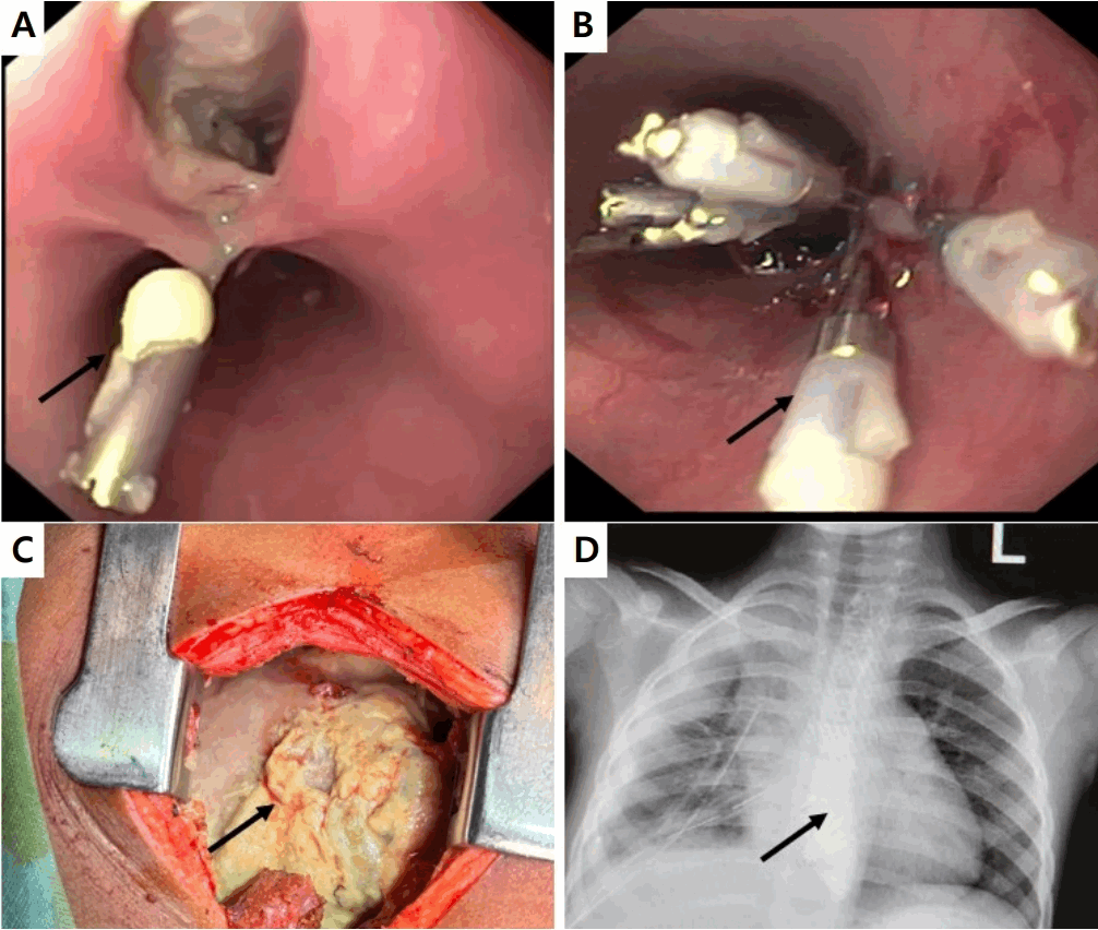
Endoscopic closure and clinical outcome of esophageal perforation with esophagopleural fistula in the index case. (A) Upper gastrointestinal endoscopy (UGIE) image showing successful application of the first Medorah hemoclip (arrow). (B) UGIE image showing application of the Olympus EZ Clip (arrow). (C) Surgical exploration image showing the purulent pleural peel (arrow). (D) Oral swallow study showing no dye leakage at 3 weeks after closure (arrow).
Typically, esophageal FB impaction occurs at anatomically narrow zones of cricopharynx, level of arch of aorta, and gastroesophageal junction (GEJ) [6]. Complications related to impacted esophageal foreign bodies include mucosal ulceration, obstruction, perforation, mediastinitis, vascular pseudoaneurysm with fistula formation and “buried treasure syndrome” [4]. Risk factors predisposing to esophageal perforation are delayed presentation, anatomical defects (tracheo-esophageal fistula repair, caustic strictures, eosinophilic esophagitis), type of foreign body (sharp objects, extravasating button battery, double magnets, water absorbent gel), and impaction in the upper cervical esophagus (predisposing to tracheal stenosis from esophageal dilation and compression) [7]. The reported complication rate following coin impaction ranges between 10%–20%, particularly when it remains impacted for more than 24 hours or with repeated unsuccessful primary instrumentation attempts, as in our case [4].
The European Society of Gastrointestinal Endoscopy guidelines (2020) recommend that endoscopic techniques play an important role in the management of esophageal perforations, with TTS and OTS clips being used for defects ≤10 mm and >10 mm, respectively [8]. OTSC systems are often preferred for larger defects because of stronger tissue compression and greater tissue capture [5]. However, the deployment cap (smallest 10 mm) and its corresponding endoscopic maximum outer diameter (14.65 mm) make them effective in children with a minimum age and weight of 5 years and 18 kg, respectively [9]. Similarly, endosuture devices and SEMS placement, which are used for larger defects (>20 mm), may be technically challenging in toddlers due to the narrow esophageal lumen and absence of smaller stent deployment systems [8]. Stents tend to fail in cervical esophagus or GEJ defects, and when the defect size is >6 cm, likely due to stent migration and ineffective tissue coverage [4]. Although ours was a very large perforation (2.5 cm), needing a total of 13 clips, previously unreported, it obviated the need for a morbid primary surgery in such a small child. Surgical intervention is needed only in extensive mediastinitis, large non-linear circumferential defects, uncontrolled sepsis, or failure of endoscopic therapy, after 4–6 weeks of the incident event, to prevent anastomotic site disruption [10].
To conclude, the present case demonstrates that multiple TTSCs may represent a feasible and effective minimally invasive option for toddlers with large linear esophageal perforation when other devices are unsuitable, depending on operator expertise, defect characteristics (≤2–2.5 cm) and timing of intervention (<24 hours) [4,5]. Defect size should ideally be estimated by direct endoscopic visualization, as contrast extravasation in CT imaging typically appears exaggerated compared to the actual defect dimensions. Impacted foreign body removal should be performed in centres with surgical backup, as early recognition and intervention may obviate surgical complications. A limitation of this study is the absence of long-term follow-up for stricture formation, especially after deploying 13 clips in a growing child. Written informed consent was obtained from the parents of the patient for the publication of this report.
Question
Which of the following is the least likely risk factor for esophageal perforation with respect to foreign body ingestion in children?
A. Sharp foreign body ingestion
B. Prior esophageal anatomical defects
C. Coin impaction in the lower esophagus less than 2 hours
D. Coin impaction anywhere in the esophagus for more than 24 hours
Answer: C. Coin impaction in the lower esophagus less than 2 hours
Supplementary material
Supplementary video clip 1 is available at https://doi.org/10.3345/cep.2026.00654.
Supplementary video clip 1 shows successful endoscopic closure of a linear lower thoracic esophageal perforation and post-coin impaction using through-the-scope clips.
Notes
Conflicts of interest
No potential conflict of interest relevant to this article was reported.
Funding
This study received no specific grant from any funding agency in the public, commercial, or not-for-profit sectors.
Acknowledgments
We are grateful to Dr. Debashish Mitra (Consultant, Pediatric Surgery), Dr. Sanghamitra Bhattacharya (Consultant, Pediatric Surgery), and Dr. Sudeshna Malakar (Consultant, Department of Clinical Imaging and Interventional Radiology) for helping us by giving their valuable inputs during the inpatient management of this case.
Author contribution
Conceptualization: SC, UCG, MKG; Data curation: NS, KC, TM; Methodology: SC, UCG, MKG; Visualization: UCG, MKG; Writing - original draft: SC, NS, KC, TM; Writing - review & editing: UCG, MKG