Prognosis of pediatric hepatic Wilson disease with ATP7B loss of function variants
Article information

Abstract
Background
Genotype-phenotype correlations in Wilson disease (WD) have so far been inconclusive.
Purpose
ATP7B variants with loss of function (LOF) may have a different trajectory. Since genotypes in Asia differ from the West, we aimed to correlate LOF variants of ATP7B with the severity and outcome of hepatic WD.
Methods
Patients with a confirmed diagnosis of WD (Leipzig criteria ≥4) were prospectively enrolled. Genetic sequencing of ATP7Bmutations was assessed by Whole-exome sequencing. For patients with variants of uncertain significance, Sanger sequencing was additionally performed on their parents to identify the inherited variants. In silico analyses were used to predict the pathogenicity of variants. Mutations that resulted in at least one truncation (nonsense, frameshift, splice site, deletions) and nontruncation (missense, synonymous) protein were defined as LOF and no LOF (NLF) respectively. Phenotypes, biochemical parameters, and outcomes were analyzed.
Results
One hundred sixteen hepatic WD children (84 boys, median age at diagnosis 8.9±3.3 years) with biallelic ATP7B mutations (62 different variants) were enrolled. The most common LOF (n=79) and NLF (n=37) variants were c.813C>A and c.3809A>G. Advanced liver disease (76% vs. 4%, P=0.004), portal hypertension (44% vs. 24%, P=0.03), neurological (39% vs. 16%, P=0.01) and renal involvement (44% vs. 13%, P=0.01) were significantly higher in LOF than in NLF. c.813C>A had higher serum exchangeable copper (6.8±4.4 μmol/L vs. 1.4±3.5 μmol/L, P=0.04) and lower disappearance of the Kayser-Fleischer ring (4% vs. 49%, P= 0.01) than c.3809A>G variants. Over a follow-up of 6.1±4.7 years, a single LOF variant did not show a poorer liver or overall outcomes in comparison to ≥2 LOF variants.
Conclusion
A single ATP7B LOF variant, especially c.813C>A was associated with advanced liver disease, portal hypertension, and extrahepatic involvement. LOF variants did not affect liver or overall outcomes.
Key message
Question: Do the loss of function (LOF) of ATP7B mutated variants affect the severity or outcomes of hepatic Wilson disease (WD)?
Finding: LOF is associated with advanced liver disease, portal hypertension, and extrahepatic involvement. LOF did not determine hepatic or overall outcomes. ATP7B: c.813C>A (truncation variant) has an aggressive course with higher predisposition to acute liver failure. Lower serum exchangeable copper levels and higher disappearance of Kayser-Fleischer ring were observed in ATP7B: c.3809A>G (nontruncation variant) suggesting a better effect of chelation and lesser systemic copper.
Meaning: Truncation of the ATP7B protein determines the severity of the phenotype in WD.

Graphical abstract. LOF, loss of function.
Introduction
Wilson disease (WD) is an autosomal recessive multisystem copper metabolism disorder caused by mutations in the ATP7B gene. The gene encodes the ATP7B protein, which belongs to the copper-transporting P-type ATPase [1]. If untreated, WD may be progressive and lethal. The disease can be kept in remission if chelation therapy is initiated on time. The hepatic variant is particularly aggressive in children, with 80% presenting with advanced liver disease and a 20% mortality rate or requiring liver transplantation [2]. Challenges in the diagnosis and delays result in irreversible severe complications and rendering treatments ineffective. There are fallacies in the interpretation of clinicobiochemical tests for WD. Genetic testing may be the closest confirmation for the diagnosis in WD. A significant variation exists in the literature regarding the prevalence of various pathogenic variants in WD. Some pathogenic variants are regional or based on race and anthropology. c.3207C>A (p.H1069Q) is common in Europe and North America, whereas c.2333G>T (p.R778L) is reported in East Asia [3,4]. Hence, it may not be appropriate to extrapolate genotype-phenotype correlations from Caucasian to Asian patients. Globally, genotype-phenotype correlations have been largely inconclusive in WD [5,6]. Families reporting rare ATP7B variants have shown phenotypic diversity among affected individuals [7,8]. In genetic disorders, mutations that result in truncated proteins are deemed to have greater severity of disease than those with intact non-functional or partially functional proteins. Nonsense and frameshift mutations lead to loss of function (LOF) due to premature termination of translation resulting in truncated proteins or degradation of the mRNA (through nonsense-mediated decay). Hence, they are generally predicted to result in severe phenotypes [9]. We aimed to correlate LOF variants of ATP7B with the severity and outcome of hepatic WD.
Methodology
Patients with a confirmed diagnosis of WD (Leipzig criteria≥4) were prospectively evaluated for genetic sequencing of ATP7B mutations. Their liver disease phenotype, extrahepatic involvement, biochemical parameters, severity of liver disease, and outcomes were analyzed.
1. Genetic testing
Genomic DNA was extracted from peripheral whole blood using a commercial DNA extraction kit (Cat. No. 51106), following the manufacturer’s protocol. DNA concentration and purity were assessed using a NanoDrop 2000 spectrophotometer (Thermo Fisher Scientific, USA), and DNA integrity was evaluated by 1% agarose gel electrophoresis. Whole-exome libraries were prepared using the Agilent SureSelect Human All Exon v8 kit (Agilent Technologies), according to the manufacturer’s instructions. Briefly, genomic DNA was fragmented into short inserts, end-repaired, and adapter-ligated. The adapter-ligated fragments were subsequently PCR-amplified to generate indexed libraries. Exonic regions were captured through hybridization of amplified libraries, followed by washing and elution of the target DNA fragments using kit-provided buffers. The enriched libraries were sequenced on an Illumina NovaSeq 6000 platform (Illumina, USA), generating 150-bp paired-end reads for downstream analysis. The Human Gene Mutation Database (HGMD) and the ClinVar database were used to confirm known pathogenic variants; variants of unknown significance (VUS) and in silico analyses were used to predict the pathogenicity of novel variants. For patients with VUS, Sanger sequencing was additionally performed on their parents to identify the inherited variants. Patients with a single heterozygous variant underwent reanalysis of raw data and/or whole-genome sequencing to detect any unidentified variants in the non-coding or deep intronic regions. The full spectrum and frequency of variants in the ATP7B gene were analyzed to identify and examine truncation and nontruncation variants.
2. Inclusion criteria
ATP7B homozygotes or compound heterozygotes with at least one pathogenic, likely pathogenic, or VUS presenting with a hepatic phenotype.
3. Exclusion criteria
Patients without ATP7B genetic sequencing, those with a single heterozygous variant, exclusive nonhepatic phenotype, and ambiguity in diagnosis.
4. Definitions
Truncation variants: nonsense (substitution in a coding base pair that results in a premature stop codon), frameshift (insertion or deletion predicted to result in a shift in the reading frame), deletions (exon deletions), and splice site (substitutions that involve a splice site). Non-truncation variants: missense (substitution in a coding base pair that results in a change of amino acid) or synonymous (substitution in a coding base pair that does not change the amino acid in a coded protein). LOF: presence of one or more truncating mutation(s). No LOF (NLF): both nontruncation mutations [10,11]. Portal hypertension: presence of gastroesophageal varices and/or hypersplenism, with or without symptoms. Standard definitions were used for compensated, decompensated, and recompensated cirrhosis [12-14]. Mild liver disease: Child-Turcotte-Pugh (CTP) score A. Advanced liver disease: CTP score B or C. Favorable liver outcome: recompensation, stable liver function, survival with the native liver. Poor liver outcome: worsening liver function, further decompensation, the need for liver transplantation, or death. Favorable overall outcome: recovery of hepatic and extrahepatic (renal, neurological, hemolysis) improvement. Poor overall outcome: new onset or progression of extrahepatic disease, neurodebilitation.
5. Ethics statement
Ethical clearance was obtained from the Institute Ethical Committee (IEC code 2022–94-EMP-128). All patients provided written consent for the study.
6. Statistical analysis
For baseline and demographic data, categorical data were represented as percentages, and continuous variables were represented as mean±standard deviation. Student t test/Mann-Whitney U test for comparison of continuous variables, and chi-square/Fisher exact test for categorical variables were used. The survival of children and the development of complications were analyzed using Kaplan-Meier survival curves. Logistic regression analysis was used to assess the effect of variables. Cox regression analysis was used to investigate the effect of variables (risk factors) on follow-up complications and mortality. Prognostic models were constructed. The discrimination of the models was described using the area under the receiver operating characteristic curve (AUROC). All AUROC models were censored. P values <0.05 were considered significant. All calculations were performed using IBM SPSS Statistics ver. 23.0 (IBM Co., USA) and R ver. 3.6 (R Foundation for Statistical Computing, Austria).
Results
1. Overall presentation
One hundred sixteen patients (84 boys) from 100 unrelated families with hepatic WD with biallelic ATP7B mutations were enrolled. The median age at diagnosis was 8.9±3.3 years. The duration of the follow-up was 6.1±4.7 years. Overall presentation included stable decompensated cirrhosis (n=50), unstable decompensated cirrhosis (n=13), compensated cirrhosis (n=18), acute liver failure (n=6), asymptomatic hypertransaminemia (n=26) and recurrent hepatitis (n=1). Two children presented with acute variceal bleeding. Fourteen siblings identified by screening had asymptomatic hypertransaminasemia (n=8) and compensated cirrhosis (n=6). Neurological involvement and renal tubular dysfunction at onset were seen in 25 and 27 children, respectively. As a departmental protocol, all patients were initiated on D-penicillamine for therapy. Trientine was started in 14 patients with D-penicillamine intolerance at follow-up. Zinc was added in 8 patients for better control of the disease. Four patients later experienced neurological worsening (D-penicillamine and trientine) and were maintained on zinc monotherapy.
2. Genotype
In total, there were 62 different variants of ATP7B; 34 missense, 13 frameshift, 11 splice site, 3 nonsense, and 1 synonymous (Fig. 1; Supplementary Table 1). Of the 116 patients, at least one LOF was seen in 79 patients. Of the 79 patients, 43 had 2 LOF. 37 patients had NLF variants. The most common variants were c.813C>A encoding p.C271X (n=55, 31%), c.3809A>G encoding p.N1270S (n=18, 10%), c.3182G >A encoding p.G1061E (n=9, 5%) and c.2906G>A encoding p.R969Q (n=6, 2%). c.813C>A and c.3809A>G comprised 70% of LOF and 49% of NLF variants, respectively. 26 novel variants (15 truncating, 11 nontruncating) were identified in 31 patients. In 114 patients, 2 affected alleles were identified. In the remaining 2 patients, there were 3 affected alleles (c.3646G>A coding p.V1216M; c.3741C>G coding p.H1247Q; c.174dup coding p.T59Hfs*19). The exon-intron location and pathogenicity of the variants are shown in Fig. 2.
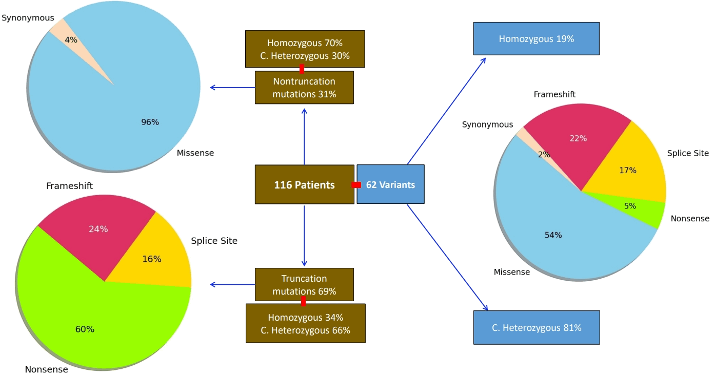
Distribution of 62 ATP7B variants in the Wilson disease cohort according to zygosity and mutation types. Variants were further classified as truncation (nonsense, frameshift, splice site) and nontruncation (missense, synonymous).

Schematic diagram highlighting the domains and pathogenicity of the various ATP7B mutations according to their exonic and intronic regions. LOF, loss of function.
3. Comparison of LOF vs. NLF
Table 1 shows the comparison of patients with LOF versus NLF. The age at diagnosis, follow-up duration, and therapy duration were similar between the 2 groups. Although CTP and pediatric end-stage liver disease (PELD) scores were similar, three-quarters of LOF and half of NLF presented with advanced liver disease. The presence of portal hypertension and extrahepatic involvement was significantly higher in LOF than in NLF. The liver and overall outcomes were not significantly different between the 2 groups.
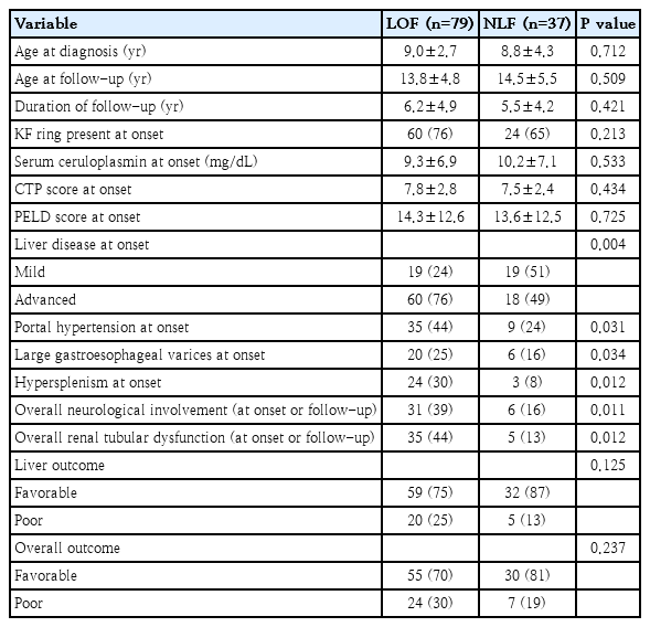
Comparison of presentation and outcomes of ATP7B LOF vs. NLF
4. Comparison of c.813C>A vs. c.3809A>G variants
c.813C>A and c.3809A>G coexisted in a compound heterozygous state in 3 patients. After exclusion of these 3 patients, the 2 mutations were compared to assess their actual contribution to the results of LOF and NLF (Table 2). c.813C>A presented with advanced liver disease and portal hypertension. Lower serum exchangeable copper levels and higher disappearance of Kayser-Fleischer (KF) ring were observed in c.3809A>G suggesting a better effect of chelation and less systemic copper. The baseline to follow-up change (Δ) in PELD score, liver outcome, and extrahepatic involvement at follow-up did not differ between the 2 groups.
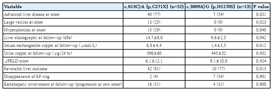
Comparison of presentation and outcomes of ATP7B variants c.813C>A versus c.3809A>G
5. Comparison of 1 vs. 2 LOF variants
Thirty-six patients with a single LOF mutation (1 LOF) were compared with 43 patients with 2 LOF mutations (≥2 LOF) as shown in Table 3. ΔPELD score, the presence of portal hypertension, and extrahepatic features were similar in both groups. The liver and overall outcomes were comparable between the 2 groups.
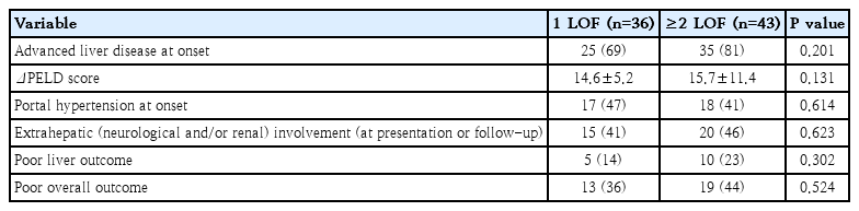
Comparison of presentation and outcomes of ATP7B 1 LOF versus ≥2 LOF variants
6. ATP7B variants in fulminant hepatic failure
Six patients presented with fulminant liver failure (FHF) with hemolysis. All 6 harbored c.813C>A. Two were homozygous; the rest were compound heterozygous with c.3809 A>G (n=2), c.2304dup encoding p.M769Hfs*26 (n=1) and c.4039G>A encoding p.G1347S (n=1). Five patients had native liver survival with therapeutic plasma exchange, while one patient died while waiting for liver transplantation.
7. Long-term outcomes
Eight patients (6 LOF, 2 NLF) with advanced liver disease died of complications. Four developed acute kidney injuries requiring renal replacement therapy, refractory encephalopathy, and pulmonary hemorrhage. The other 4 developed overwhelming septicemia and multiorgan dysfunction. Four patients (all LOF) with severe hypersplenism required partial splenic artery embolization to improve blood counts for the sustenance of chelation. Three patients (2 unstable decompensated cirrhosis, 1 recurrent bleeding gastric varices) underwent liver transplantation. Nine patients (5 LOF, 2 NLF) with worsening liver function were waitlisted for liver transplantation until the last follow-up. Fig. 3 shows the Kaplan-Meier survival curves of the 3 comparison groups (LOF vs NLF; c.813C>A vs. c.3809A >G, 1 LOF vs. ≥2 LOF). Although ≥2 LOF was observed to have poorer liver and overall outcomes than 1 LOF, it was not statistically significant. 12% of the cohort had issues with drug compliance. Supplementary Figs. 1 and 2 depict the outcomes of the LOF and NLF variants in the Sankey diagrams.

Kaplan-Meier curve analysis showing clinical outcomes according to ATP7B mutations. (A) truncated versus nontruncated mutations (log rank test, P=0.18). (B) c.813C>A versus c.3809A>G (log rank test, P=0.27). (C) 1 LOF vs. ≥2 LOF (log rank test, P=0.38).
Discussion
Despite WD being a monogenic disease, there is variable expressivity and phenotypic diversity among affected individuals at different time points, within families, between siblings, and across different ethnicities [7,8]. The reasons are unknown. Phenotype may depend on a number of factors, such as delayed diagnosis, pitfalls in diagnostic criteria, and adherence to therapy. Clinicians attempt to unravel genotype-phenotype correlations to predict the natural history and outcomes of the probands and their first-degree relatives. In this study, we explored whether LOF of ATP7B variants could correlate with the hepatic phenotypes of WD.
The known pan-India variants, c.813C>A (p.C271X), c.3809A >G (p.N1270S) and c.3182G>A (p.G1061E) represent 46% of our genetic cohort [15]. ATP7B c.813C>A is unique to South Asian ancestry and its migrants in Western countries. ATP7B c.3809A>G and c.3182G>A have been reported globally. The types of mutations in our cohort (missense [45%], nonsense [32%], frameshift [13%], and splice site [8%]) matched the worldwide HGMD (missense/nonsense [62%], frameshift [26%], and splice site [8%]) [4]. Of the 62 variants reported, 81% were in a compound heterozygous state, and 68% were deemed LOF based on the truncated proteins.
Variant c.813C>A which contributed to a large proportion of LOF, was associated with more advanced liver disease and portal hypertension than NLF. However, genetics did not contribute to liver outcomes, as improvements in the PELD score were similar between the LOF versus NLF and c.813C>A versus c.3809A>G groups. The PELD score does not include the impact of fibrosis and portal hypertension. In our cohort, liver elastography at follow-up was lower in the NLF than in the LOF, showing some improvement in liver architecture. Portal hypertension in WD is a standalone factor for worse transplant-free survival, especially if liver stiffness is >15 kPa [16]. All the patients presenting with FHF had the presence of one or more c.813C>A. As with our previous experience, we have shown that the time lag from the onset of symptoms to diagnosis is approximately 2 months, even in patients with severe liver disease [2]. Lack of early identification and poor referral patterns may not be the sole reasons. Early decompensation raises questions about the in vivo functional aspects of the p.C271X formed from the c.813C>A variant with regard to parenchymal injury and fibrosis. Is this variant the reason for the aggressive nature of pediatric hepatic WD? Functional studies and in silico tests are required to understand the behavior of this variant.
There are conflicting results regarding the genotype-phenotype correlation in WD. Ferenci et al. [6] studied 394 mutations, of which c.3207A>G was the most common mutation. The authors were unable to demonstrate any correlation between the hepatic or neurological phenotypes in the predominantly Caucasian cohort. In contrast, our study showed that LOF had higher neurological and renal involvement than NLF. Pop et al. [17] observed that in children with liver failure and hemolytic anemia, c.2817G >T and c.2530A>T variants were more frequent than less severe variants such as c.3207A>G. Studies in the past have shown that LOF was associated with early onset of disease, lower ceruloplasmin, higher copper, and a higher prevalence of FHF [6,9,11,18-20]. We did not find any correlation between the mutations and serum ceruloplasmin levels.
Due to the inefficiency of the defective ATP7B protein, the amount of copper handled by the liver and released into the circulation will also depend on its residual function. It is expected that proteins from nontruncation mutations should function better than those from truncation mutations. Serum exchangeable copper is a labile form of copper that is deposited into receptive organs (brain, kidney, cornea). Our group has previously shown that serum exchangeable copper is a unique method to assess the long-term adequacy of chelation, now incorporated in the EASL (European Association for the Study of the Liver) guidelines [21,22]. In the present study, the c.813C>A variant had significantly higher serum exchangeable copper levels than the c.3809A>G variant, although neither group had fully achieved optimal chelation. Indirect evidence of chelation was also observed from the disappearance of the KF ring, higher in c.3809A>G than c.813C>A variant. The efficacy of chelation with c.813C>A variant needs to be evaluated through pharmacokinetic studies in the future.
One would expect a linear correlation between the number of LOF and the severity of the disease. Nayagam et al. [10] observed that there was worse transplant-free survival for patients with at least 1 LOF variant on chelation therapy. However, LOF did not distinguish between FHF and chronic liver disease phenotypes in their study. We could not demonstrate a worse liver outcome with ≥2 LOF compared with 1 LOF. We agree with Nayagam et al. that 1 LOF is probably enough to sufficiently lower ATP7B function below the threshold and cause damage. The second LOF adds little to the impact. Liver recovery depends on many factors beyond genetics. Since WD is a multisystemic disorder, it is pragmatic to take overall recovery into consideration. Over time, the predominant phenotype of Wilson's disease may shift to affect a different organ. How the body handles the copper reservoirs is an enigma, typically observed with the age-phenotype effect or the sex-phenotype effect [6,23]. We have demonstrated that extrahepatic manifestations at presentation and follow-up are higher with LOF than with NLF variants, more so with c.813C>A variant. However, due to multifactorial reasons, the overall recovery was similar between the 2 groups. Availability of equitable health care, affordability of drugs, referral systems, awareness programs, and geographical inaccessibility influence outcomes in developing nations that struggle with rare genetic diseases. There is a lack of multispecialty expertise to manage WD in the country; hence, some of the phenotypes are poorly managed. Indolent and occult manifestations, such as renal tubular dysfunction and bone damage, are often missed at diagnosis and not considered during follow-up due to a lack of vigilance. Of the multiple factors that influence the severity of the disease, the most important are the degree of liver fibrosis and copper burden. Truncating ATP7B variants may result in greater loss of protein function and inability of the defective ceruloplasmin to carry copper atoms. This results in higher intracellular copper, potentially contributing to more severe hepatic phenotypes and fibrosis progression. Excess intrahepatic copper then induces oxidative stress and mitochondrial dysfunction, leading to hepatocyte apoptosis and necrosis [24]. This injury activates profibrogenic pathways, resulting in hepatic stellate cell activation and progressive extracellular matrix deposition [25,26]. Additionally, modifier genes, epigenetic alterations, immune dysregulation, and altered cellular signaling pathways may influence the function of ATP7B and contribute significantly to severity of disease [25,27].
One of the fallacies in the interpretation of genotype-phenotype results worldwide, including ours, is the assumption that the missense mutation is nontruncating and that milder phenotypes are anticipated. While truncating defects cause a definite loss of protein, missense mutations may cause a variety of protein aberrations, such as misfolding, loss of expression, copper-binding issues, and disrupted protein trafficking [28]. Hence, the result of the “missense dysfunctional protein” may be as deleterious as the LOF. Until functional assays are performed on the missense variants, the true genotype-phenotype correlation will always remain in limbo. Calvo et al. [29] screened 101 variants of ATP7B for expression and copper transport activity in human fibroblasts that lacked active ATP-dependent copper transporters. They identified that 3 variants (p.S657R, p.G1061E, and p.G1266R) resulted in the complete inactivation of copper transport. This is relevant as p.G1061E (c.3182G>A) was also seen in 2% of our cohort. With collaborative data, it needs to be determined whether this mutation results in severe disease compared to the rest. Meticulously performing functional assays on over 1300+ ATP7B globally known variants is cumbersome and cost-prohibitive, as the final endpoint is chelation therapy, irrespective of the variant. However, functional assays could open doors to new therapeutic targets such as variant-specific chaperones. Another alternative in the future is the correlation of the missense mutation with the quantification of the ATP7B peptide, which yields in 94% of genetically ambiguous cases [30]. The ultimate bridge in understanding the genotype and phenotype is the use of transcriptomics, which will assess the dynamics of the defective protein in the target organs. The ATP7B gene may also be governed by epigenetic factors and modifier genes; hence, there may be discordances in the direct genotype-phenotypic variations. There is limited and speculative data on the same presently [31].
In conclusion, a single LOF variant in ATP7B was associated with advanced liver disease, portal hypertension, and extrahepatic involvement but did not affect the liver or overall outcomes. Variant c.813C>A may have an aggressive course in WD.
Supplementary materials
Supplementary Figs. 1-2 and Supplementary Table 1 are available at https://doi.org/10.3345/cep.2026.00689.
Sankey diagram illustrating the phenotype distributions and clinical outcomes associated with ATP7B loss of function variants.
Sankey diagram illustrating the phenotype distributions and clinical outcomes associated with ATP7B no loss of function variants.
In silico prediction of the functional impact of ATP7B variants based on their location and pathogenicity
Notes
Conflicts of interest
No potential conflict of interest relevant to this article was reported.
Funding
Extramural research grant was provided by the Department of Health Research, Indian Council of Medical Research, New Delhi (R. 11014/50/2023-GIA/HR) DHR.
Acknowledgments
Department of Science and Technology (INSPIRE Fellowship) for supporting the PhD thesis program (IF230357).
Author contribution
Conceptualization: MSS; Data curation: AKM, MSS, AD, PG, AM; Formal analysis: AKM, MSS; Funding acquisition: MSS; Methodology: AKM, MSS, AM; Project administration: AKM, MSS, AD, PG, AM; Visualization: AKM, MSS, AS; Writing - original draft: AKM; Writing - review & editing: AKM, MSS