A rare genetically confirmed case of apparent mineralocorticoid excess syndrome due to a homozygous HSD11B2 variant in a 2-year-old child with severe hypertension: response to targeted therapy
Article information


Graphical abstract. Apparent mineralocorticoid excess syndrome. ACTH, adrenocorticotropic hormone; PTH, parathyroid hormone; US, ultrasonography; RI, resistive index; CTA, computed tomography angiography; BP, blood pressure.
Apparent mineralocorticoid excess (AME) is a rare autosomal recessive form of hypertension caused by dysfunction of the 11β-hydroxysteroid dehydrogenase type 2 (11β-HSD2) enzyme. Mutation in the 11β-hydroxysteroid dehydrogenase type 2 HSD11B2 gene located on chromosome 16q22 [1,2]. 11HSD-2 is expressed in principal cells of the collecting ducts. Activity loss of 11β-HSD2 leads to continuous cortisol accumulation, overstimulation of the mineralocorticoid receptor, increased sodium reabsorption, and potassium loss. Clinical characterization includes early-onset hypertension, hypokalemia, metabolic alkalosis, suppressed plasma renin activity, and absence of circulating aldosterone [3,4]. AME is classified into types I and II [1].
A 2-year-old girl was referred for evaluation of hypokalemia identified during admission for acute tonsillitis. The patient had a 3-day history of fever, upper respiratory symptoms, poor oral intake, and reduced activity. Her fever reached 38°C at home and was accompanied by cough and decreased feeding. Initially diagnosed with acute tonsillitis and treated with oral antibiotics, the patient presented to the emergency department due to persistent symptoms.
Her medical history included admission at 16 months for fever, vomiting, and diarrhea, during which hypokalemia (2.5 mmol/L) was noted but attributed to gastrointestinal losses, with no further intervention. At 22 months, she was admitted after accidental clorox ingestion and again found to have hypokalemia (2.5 mmol/L), which was not further investigated.
Antenatal history was unremarkable. The patient was born at term (36 weeks and 6 days' gestation) and required neonatal intensive care unit admission for respiratory distress, and was small for gestational age at 1.97 kg. The parents are second-degree relatives, and she has a healthy 10-month-old sister.
On examination, growth parameters revealed a weight of 9.6 kg (5th percentile) and a height of 85 cm (25th percentile) [2]. Vital signs were stable, with a blood pressure (BP) of 95/58 mmHg and a peripheral oxygen saturation of 96%. Dysmorphic features included brittle hair, flat nasal bridge, large forehead, frontal bossing, long eyelashes, and ear tag. Patient was active, conscious, well hydrated, and perfused. Initial laboratory investigations are shown in Table 1.
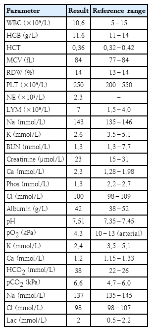
Summary of clinical, biochemical, hormonal, imaging, and genetic findings
On the second day of admission, the nephrology team was consulted due to persistent hypokalemia requiring repeated potassium chloride (KCl) supplementation. Initial management included monitoring intake and output, assessment of renin and aldosterone levels, confirmation of normal urine electrolytes, renal ultrasound, initiation of oral KCl 5 mmol once daily, dietary review, and genetic evaluation. The patient was polyuric, with urine output of 6–8 mL/kg/hr. Kidney ultrasound revealed bilaterally enlarged echogenic kidneys with preserved corticomedullary differentiation, medullary nephrocalcinosis, and multiple small bilateral cysts [5]. Despite intervention, hypokalemia persisted, requiring gradual increases in KCl.
BP progressively increased during hospitalization, reaching 136/97 mmHg, exceeding the 95th percentile for age [6]. The patient required pro re nata hydralazine followed by oral hydralazine, which was titrated to the maximum tolerated dose, and atenolol was added, low renin (1 mIU/L) and low aldosterone (<26 pmol/L), consistent with mineralocorticoid excess. These findings, together with persistent hypokalemia, metabolic alkalosis, and hypertension, were highly suggestive of AME syndrome.
Ultrasound Doppler of the renal arteries showed elevated bilateral renal resistive indices and features of renal parenchymal disease, including nephromegaly, medullary nephrocalcinosis, small renal cysts, and cortical calcification. Computed tomography angiography (CTA) showed no renal artery stenosis. Due to dysmorphic features, whole exome sequencing was performed and identified a homozygous pathogenic HSD11B2 variant (c.622C>T; p.Arg208Cys), confirming AME syndrome. An incidental heterozygous pathogenic UBE3B variant (c.518C>A; p.Ser173*) was detected but was not clinically relevant.
Management included spironolactone 5 mg orally once daily with continued oral and intravenous (IV) KCl. As spironolactone was gradually increased to 8 mg/kg/day, potassium improved to 3.1 mmol/L and IV supplementation was discontinued. Oral KCl was gradually tapered and stopped once serum potassium stabilized at 3.7–4.1 mmol/L. Hydrochlorothiazide was briefly introduced but discontinued after 5 days due to worsening hypokalemia and no significant BP improvement. Spironolactone, hydralazine, and atenolol ultimately achieved BP control, improving from a peak of 136/97 mmHg to 100/60–110/70 mmHg range (≈50th percentile) [6], by discharge. The patient was discharged clinically stable on spironolactone 8 mg/kg/day, atenolol 5 mg twice daily, and hydralazine 25 mg three times daily.
AME is a rare autosomal recessive disorder caused by mutations in the HSD11B2 gene, which encodes the 11β-HSD2 enzyme that converts cortisol to cortisone [3,4]. When this enzyme is deficient, excess cortisol binds to mineralocorticoid receptors, leading to sodium retention, potassium loss, hypertension, and metabolic alkalosis.
Classic AME typically presents in infancy or early childhood with severe hypokalemia, polyuria, polydipsia, poor growth, and early-onset hypertension. Milder forms may present later with less pronounced symptoms and lower BP [1]. The patient in this report, who presented at age 2 with severe hypokalemia, polyuria, dysmorphic features, and increasing BP, is consistent with the classic AME phenotype.
Lab tests showed low renin and aldosterone, high cortisol, and a high cortisol-to-cortisone ratio, suggesting mineralocorticoid receptor overactivity despite low aldosterone. Imaging showed bilateral nephrocalcinosis, cysts, and high resistive indices, suggesting chronic kidney changes from excessive mineralocorticoid activity. A CTA ruled out renal vascular obstruction, confirming a hormonal cause.
Whole exome sequencing found a homozygous pathogenic variant in HSD11B2 (p.Arg208Cys), confirming AME. This case highlights the importance of considering AME in children with early, resistant hypokalemia and hypertension, especially with parental consanguinity. The patient’s dysmorphic features prompted genetic testing, which confirmed the diagnosis and guided treatment.
The main treatment goals in AME are correcting hypokalemia and controlling hypertension. Mineralocorticoid receptor blockers like spironolactone are the main treatment and stabilized this patient’s potassium and BP. Potassium supplementation was initially required but later discontinued after spironolactone was titrated to 8 mg/kg/day. Thiazide diuretics may reduce hypercalciuria but can worsen hypokalemia and require close monitoring [5-7]. Additional BP medications, including hydralazine and atenolol, were also needed. Long-term care includes salt restriction, continued mineralocorticoid receptor blockade, and kidney monitoring. Kidney transplant can cure severe refractory cases but is rarely required in classic AME [7-9].
This case highlights the importance of early recognition of AME in patients with recurrent hypokalemia and hypertension. It also emphasizes a multidisciplinary approach involving nephrology, genetics, and nutrition. Combining clinical, laboratory, imaging, and genetic findings was essential for diagnosis and treatment. Classic and nonclassic AME are compared in Fig. 1.
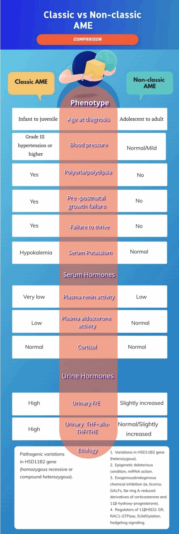
Comparison between classic and nonclassic AME, highlighting phenotypic, biochemical, and etiological differences.1) The figure was created by the authors using a licensed design platform. AME, apparent mineralocorticoid excess; F/E, free cortisol to free cortisone ratio; THF, tetrahydrocortisol; THE, tetrahydrocortisone; miRNA, microRNA; GALFs, glycyrrhetinic acid-like factors; GR, glucocorticoid receptor; RAC1, ras-related C3 botulinum toxin substrate 1; SUMOylation, small ubiquitin-like modifier proteins conjugation.
In summary, this rare, genetically confirmed case of AME reveals that early diagnosis and targeted treatment can improve growth, correct electrolyte imbalance, and control BP, reducing the risk of long-term kidney damage. AME should be considered in children with persistent hypokalemia and hypertension, especially in consanguineous populations.
Timely diagnosis of AME is essential, as untreated hypokalemia and hypertension can cause significant organ damage, including early stroke, cardiac complications, ocular disease, and progressive renal dysfunction. The clinical presentation of AME is variable, and hypertension may be absent initially, which can delay diagnosis. Early recognition through comprehensive clinical assessment, laboratory evaluation, and genetic confirmation, combined with appropriate management, is crucial to prevent long-term complications and improve outcomes.
Question
Which combination of clinical and laboratory findings most strongly suggests apparent mineralocorticoid excess syndrome in a child with persistent hypokalemia?
A. High renin and hyperkalemia
B. Low renin and low aldosterone
C. Hypercalcemia and hypotension
D. Normal electrolytes with proteinuria
Answer: B
Notes
Conflicts of interest
No potential conflict of interest relevant to this article was reported.
Funding
This study received no specific grant from any funding agency in the public, commercial, or not-for-profit sectors.
Ethics statement
The study was conducted in accordance with the Declaration of Helsinki. Ethical approval was obtained from the local Research Ethics Committee of Scientific Research Center of Prince Sultan Military Medical City. (IRB Approval No: E-2841; approved on 2 March 2026). Written informed consent was obtained from the patient’s parents prior to participation. Written informed consent for publication of this case report and accompanying images was obtained from the patient’s legal guardians.
Author contribution
Conceptualization: BA, ME, GA, GA; Data analysis: BA, ME, GA, GA. Writing-original draft preparation: BA, ME, GA, GA; Writing-review and editing: BA, ME, GA, GA.