Patient-independent automated pediatric seizure monitoring based on expert-labeled electrographic ictal data as core reference knowledge
Article information

Abstract
Background
Automated seizure detection using scalp electroencephalography (EEG) is essential to the efficient monitoring of seizures in patients with epilepsy. However, patient-independent seizure detection remains challenging, primarily because of the inherent intersubject variability in EEG characteristics.
Purpose
We proposed a patient-independent seizure detection approach based on 1,604 single-channel electrographic focal-onset ictal EEG segments verified by epileptologists in patients with focal epilepsy.
Methods
We constructed deep learning models trained on these segments and applied them to individual EEG channels to identify seizure occurrences. To evaluate patient-independent detection performance, we conducted internal validation using the 2 datasets employed for segment acquisition, followed by external validation with an independent unseen dataset obtained from a tertiary medical institution.
Results
In the internal validation using a leave-one-patient-out scenario, overall sensitivity, false alarm rate, and latency were 80.1%–100%, 0.64–1.13/hr, and 8.0–21.4 seconds, respectively. In the external validation using the final seizure detection model, the corresponding values were 100%, 0.38–0.67/hr, and 10.0–10.2 seconds, respectively, according to detection length. This technique outperformed those of previous patient-independent studies that employed relatively simple deep learning tasks.
Conclusion
A curated set of expert-labeled ictal EEG segments served as the core reference knowledge for the proposed seizure detector in recognizing seizure occurrence in unseen patients. Because the proposed approach analyzes individual channels in parallel, it may be clinically applicable to continuous seizure monitoring, particularly in wearable seizure detection systems with a limited number of channels.
Key message
Question: Can deep learning models trained on expert-verified electroencephalography segments reliably detect seizures and be applied to single-channel wearable monitoring?
Finding: This technique demonstrated 80.1%–100% sensitivity and low false-alarm rates. On 1,604 expert-labeled segments, the model outperformed previous studies in terms of latency and accuracy across unseen datasets.
Meaning: Expert-labeled data are essential for achieving universal seizure detection. This channel-specific approach enabled efficient and continuous monitoring using wearable devices.
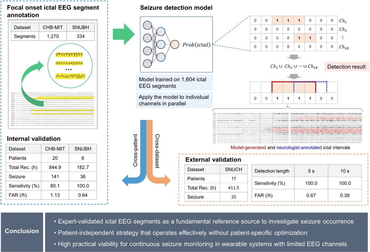
Graphical abstract. EEG, electroencephalography; CHB-MIT, Children’s Hospital Boston-Massachusetts Institute of Technology; SNUBH, Seoul National University Bundang Hospital; FAR, false alarm rate.
Introduction
Electroencephalography (EEG) is a primary tool for diagnosing epilepsy, as it plays a key role in recording the brain’s electrical activity during clinical seizures [1,2]. However, continuous EEG monitoring is labor-intensive, which has prompted extensive research into machine learning and deep learning approaches for automated seizure detection [3-5]. In particular, automated seizure detection using scalp EEG has been shown to reduce clinical workload [6] and to support wearable applications in out-of-hospital settings [7,8].
Two main strategies known as patient-specific and patient-independent models have been proposed for automated seizure detection [6]. Patient-specific models generally demonstrate superior performance because they are trained on data from a single individual. However, this approach limits clinical utility, as each model is applicable only to the patient who provides the training data. In contrast, once adequately trained, patient-independent models can be applied across multiple patients—including those who cannot provide labeled data—making them more practical and scalable [9].
Recently, deep learning approaches have advanced patient-independent seizure detection using scalp EEG. To evaluate generalizability, many studies have adopted cross-patient [10-13] or cross-dataset [9,14] validation methods. Nonetheless, performance remains limited by substantial intersubject variability in seizure patterns on EEG. This limitation is particularly evident in focal epilepsy, in which seizure characteristics vary according to ictal onset zones [15], and even within a single patient during prolonged or spatially spreading seizures [16]. To address intersubject variability, diverse ictal EEG patterns should be incorporated into a reference dataset, and their generalizability should be validated using external cohorts [17,18].
To address the limited generalizability of existing seizure detection models, this study aimed to design a deep learning-based, patient-independent model trained on 1,604 focal-onset ictal EEG segments annotated from 35 patients in public and in-house EEG datasets to enable automated detection of focal seizures. The model’s generalizability was further assessed through external validation.
Methods
1. Dataset
We used scalp EEG recordings from (1) 23 patients in the Children’s Hospital Boston-Massachusetts Institute of Technology (CHB-MIT) scalp EEG database (mean age± standard deviation, 10.0±5.7 years; 18 females and 5 males; 169 seizures; 956.6 hours of recording; chb24 excluded because of absence seizures) and (2) 12 patients with focal epilepsy from Seoul National University Bundang Hospital (SNUBH) (10.1±5.1 years; 2 females and 10 males; 91 seizures; 290.9 hours) for focal-onset ictal EEG segment annotation, model construction, and internal validation; these were referred to as the CHB-MIT and SNUBH datasets, respectively. Additional scalp EEG recordings from 11 patients with focal epilepsy at Seoul National University Children’s Hospital (SNUCH) (14.7±6.9 years; 6 females and 5 males; 25 seizures; 411.5 hours) were used for external validation and were referred to as the SNUCH dataset.
A longitudinal bipolar montage with 18 channels—Fp1–F3, F3–C3, C3–P3, P3–O1, Fp2–F4, F4–C4, C4–P4, P4–O2, Fp1–F7, F7–T3 (F7–T7), T3–T5 (T7–P7), T5–O1 (P7–O1), Fp2–F8, F8–T4 (F8–T8), T4–T6 (T8–P8), T6–O2 (P8–O2), Fz–Cz, and Cz–Pz—was used in accordance with the international 10–20 system (channels in parentheses correspond to the CHB-MIT dataset). Two epileptologists (JC and HK) annotated ictal intervals in the EEG recordings of the SNUBH and SNUCH datasets using video-EEG monitoring. They reviewed ictal EEG recordings based on the publicly available annotations of the CHB-MIT dataset and reannotated ictal intervals to ensure distinct morphological characteristics as much as possible. The institutional review board of SNUBH approved this study (No. B-2503-963-108) and waived the requirement for informed consent because of the retrospective nature of the study. This study was conducted in accordance with the principles of the Declaration of Helsinki. Fig. 1 illustrates the overall process of the study.
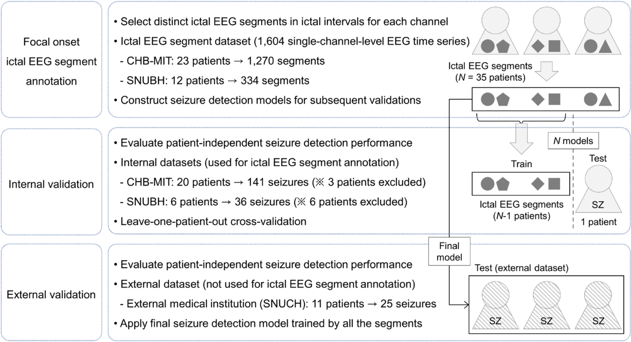
Overall study process. Two epileptologists annotated distinct electrographic focal-onset ictal EEG segments in ictal intervals for each channel, yielding an ictal EEG segment dataset comprising 1,604 singlechannel- level EEG time series. Seizure detection models were constructed by the ictal EEG dataset to evaluate patient-independent seizure detection performance using the following steps. In the internal validation, the assessment was conducted of 2 internal datasets (CHB-MIT and SNUBH) used for the ictal EEG segment annotation by the leave-one-patient-out scenario. In the external validation, the assessment was conducted for an external dataset (SNUCH) not used for the ictal EEG segment annotation by our final seizure detection model. CHB-MIT, Children’s Hospital Boston-Massachusetts Institute of Technology; EEG, encephalography; SNUBH, Seoul National University Bundang Hospital; SNUCH, Seoul National University Children’s Hospital.
2. Focal-onset Ictal EEG segment annotation
The primary objective of focal-onset ictal EEG segment annotation was to collect as many electrographic seizure patterns as possible within ictal intervals that were clearly identifiable in individual channels from multiple patients with focal epilepsy. The 2 epileptologists performed ictal EEG segment annotation as follows: (1) they reviewed each ictal interval in the CHB-MIT and SNUBH datasets; (2) examined ictal EEG segments for each channel within each ictal interval; and (3) annotated the time points corresponding to the onset and offset of ictal EEG segments in which electrographic morphological changes were clearly discernible in each channel. They finally selected the ictal segments that both of them fully agreed with each other. Through these annotation steps, we obtained a focal-onset ictal EEG segment dataset comprising single-channel EEG time series from 35 patients with focal epilepsy. This dataset was used to construct deep learning-based seizure detection models for subsequent event-level evaluation.
3. Internal validation
The primary objective of the internal validation was to investigate patient-independent seizure detection performance using the 2 internal datasets (CHB-MIT and SNUBH), which were used for ictal EEG segment annotation, through leave-one-patient-out cross-validation to eliminate the bias associated with training and evaluating on the same patient. Among the 35 patients in the internal datasets, ictal EEG segments from 34 patients were used to train a seizure detection model, and ictal intervals from the remaining patient were used for event-level evaluation of seizure detection performance in each round. For instance, the segments from chb01 were excluded when testing the chb01’s ictal intervals. Because the ultimate aim was to detect seizures rather than individual ictal EEG segments, ictal intervals for each patient were selected as the target seizure events to be detected by the models.
Considering the recently reported duration of focal seizures [19], 3 patients (chb06, chb14, and chb16) from the CHB-MIT dataset and 6 patients (SNUBH02, SNUBH04, SNUBH06, SNUBH07, SNUBH08, and SNUBH11) from the SNUBH dataset were excluded from event-level evaluation because their mean seizure durations were less than 30 seconds. However, their ictal EEG segments were included for model construction because ictal EEG segments were not the target events in event-level evaluation. Therefore, event-level evaluation was performed for 26 patients (20 from the CHB-MIT dataset and 6 from the SNUBH dataset).
4. External validation
The objective of the external validation was to investigate patient-independent seizure detection performance using an external dataset (SNUCH) that was not used for ictal EEG segment annotation, by applying the final seizure detection model. All EEG recordings from the 11 patients in the external dataset were obtained at a tertiary medical institution that was completely independent of ictal EEG segment annotation and model construction. The final seizure detection model was trained on all ictal EEG segments from the 35 patients in the internal datasets. Event-level evaluation was then performed for the 11 patients by applying the model to the entire EEG recordings of individual channels in parallel, using the same postprocessing steps and evaluation metrics as in the internal validation. To investigate the effect of detection length (L) on performance, external validation was additionally conducted with L = 10 seconds.
To build seizure detection models, we utilized a stacked 1-dimensional (1D) convolutional neural network (CNN) architecture with a 2-second EEG time series as an input signal and an ictal probability (0 to 1, ictal ≥0.5) as a model output, adopted from previous studies [20,21]. The architecture had 2 CNN modules in parallel that each one had a filter size of 1×3 and 1×5, respectively, to extract multi-scale temporal features.
Supplementary methods provide detailed information on preprocessing, postprocessing (as shown in Supplementary Fig. 1), evaluation metrics, feature visualization, and model architecture (as shown in Supplementary Fig. 2).
Results
1. Focal-onset ictal EEG segment annotation
We obtained 1,270 and 334 ictal EEG segments from 23 patients in the CHB-MIT dataset and 12 patients in the SNUBH dataset, respectively. In the CHB-MIT dataset, the number of ictal EEG segments per patient ranged from 6 to 207 (mean±standard deviation, 55.2±45.5), and the segment lengths ranged from 4.6±1.2 to 16.3±14.6 seconds (overall mean±standard deviation, 10.5±8.1 seconds). In the SNUBH dataset, the number of ictal EEG segments per patient ranged from 5 to 51 (27.8±14.1), and the segment lengths ranged from 4.0±1.7 to 16.3±19.3 seconds (9.1±8.2 seconds). Supplementary Tables 1 and 2 present detailed patient information for focal-onset ictal EEG segment annotation in the CHB-MIT and SNUBH datasets, respectively. Supplementary Figs. 3 and 4 illustrate representative focal-onset ictal EEG segments annotated by epileptologists in the CHB-MIT and SNUBH datasets, respectively.
2. Internal validation
Overall patient-independent seizure detection performance showed a sensitivity, false alarm rate (FAR), and latency of 80.1% (95% confidence interval [CI], 72.7–85.8), 1.13/hr (1.03–1.24), and 8.0±8.8 seconds (95% CI, 4.2–11.8), respectively, for the CHB-MIT dataset; and 100%, 0.64/hr (0.53–0.77), and 21.4±32.6 seconds (95% CI, 3.5–47.8), respectively, for the SNUBH dataset, based on leaveone-patient-out cross-validation. Overall sensitivity and FAR were defined as the total number of true positives (TPs) divided by the total number of true seizures, and the total number of false alarms (FAs) divided by the total recording time of all patients, respectively, in each dataset. Overall latency was calculated as the mean latency across all patients in each dataset.
In the CHB-MIT dataset, 113 of 141 seizures were successfully detected, and 953 FAs were observed during a total recording time of 844.9 hours. In the SNUBH dataset, all 36 seizures were successfully detected, and 117 FAs were observed during a total recording time of 182.7 hours. Supplementary Tables 3 and 4 present detailed results of the internal validation for the CHB-MIT and SNUBH datasets, respectively.
3. External validation
Overall patient-independent seizure detection performance demonstrated a sensitivity, FAR, and latency of 100%, 0.67/hr (0.59–0.75), and 10.0±19.8 seconds (0.2–22.6), respectively, for a detection length (L) of 5 seconds; and 100%, 0.38/hr (0.32–0.44), and 10.2±19.8 seconds (0.2–22.2), respectively, for L = 10 seconds, based on the final seizure detection model. All 25 seizures were successfully detected regardless of L, with highly similar latencies. However, 276 and 155 FAs were observed during a total recording time of 411.5 hours for L = 5 seconds and L = 10 seconds, respectively. Table 1 presents detailed results of the external validation for the SNUCH dataset. Fig. 2 illustrates a representative TP result from the eventlevel evaluation corresponding to the first seizure of SNUCH_E05 (a rescaled time-axis version is shown in Supplementary Fig. 5).
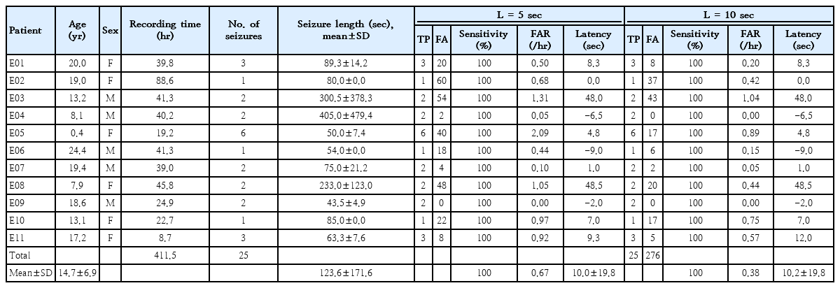
Patient information and seizure detection performance of external validation of data for 11 patients from Seoul National University Children’s Hospital dataset according to detection length (L)

A representative true-positive result in the event-level evaluation corresponding to the first seizure for SNUCH_E05. The blue- and red-colored vertical dashed lines represent epileptologist-annotated (58 seconds) and model-generated (47 seconds) ictal intervals, respectively. The upper panel represents the EEG time series with distinct seizure characteristics, particularly in the left hemisphere (red-colored channel names and signals). The lower panel represents post-processed outcomes: the blue- and red-colored solid lines represent epileptologist-annotated and channel-wise model-generated ictal intervals, respectively. The gray-colored solid lines represent ictal probabilities for individual channels. EEG, electroencephalography; SNUCH, Seoul National University Children’s Hospital.
Fig. 3 presents the ictal and interictal feature distributions for 4 channels of SNUCH_E05. Each channel included 582 ictal and 3,500 interictal windows. The center-to-center distances and Kullback-Leibler (KL) divergence values were 6.91, 3.13, 5.97, and 5.65; and 3.37, 0.84, 4.20, and 1.16 for C3–P3, C4–P4, T3–T5, and T4–T6, respectively. The KL divergence values for C3–P3 and T3–T5 were approximately 4.0 and 3.6 times higher than those for C4–P4 and T4–T6, respectively.
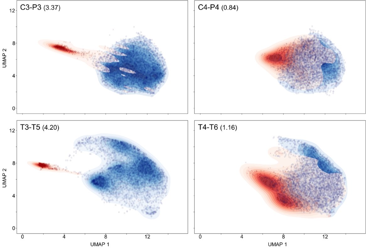
Visualization of feature vectors from the last convolutional layer based on UMAP for the 4 channels (C3–P3, C4– P4, T3–T5, and, T4–T6) of the SNUCH_E05. The red-colored cross marks and blue-colored circles represent the ictal and interictal windows, respectively. The red- and blue-colored regions represent ictal and interictal distributions, respectively, based on Gaussian kernel density estimation. The darker the color, the higher the density. The numbers in parentheses represent Kullback-Leibler divergence values between the ictal and interictal distributions. SNUCH, Seoul National University Children’s Hospital; UMAP, uniform manifold approximation and projection.
Discussion
We demonstrated a patient-independent seizure detection approach using deep learning models trained on 1,604 single-channel focal-onset ictal EEG segments derived from long-term scalp EEG recordings of patients with focal epilepsy. Overall sensitivity, FAR, and latency were 80.1%–100%, 0.64–1.13/hr, and 8.0–21.4 seconds, respectively, in the internal validation based on a leave-onepatient-out scenario; and 100%, 0.38–0.67/hr, and 10.0–10.2 seconds, respectively, in the external validation using the final seizure detection model, depending on detection length. Expert-labeled electrographic ictal EEG segments may facilitate seizure detection in unseen patients with focal epilepsy in a patient-independent manner, underscoring the potential clinical utility of this approach in settings requiring continuous automated seizure monitoring without patient-specific pretraining.
1. Seizure detection based on Ictal EEG segments
Most recent deep learning studies on patient-independent seizure detection have relied on annotated ictal intervals from multichannel EEG recordings as training datasets for seizure detection models [9-12]. However, because focal seizures exhibit polymorphic EEG characteristics and propagate from their onset zones to other channels as they progress [15,16], multichannel ictal data tend to be highly heterogeneous, particularly when derived from EEG recordings of focal seizures. Moreover, some data may contain ictal properties in only a subset of channels when seizures are localized to a specific region. In addition, intersubject variability has been recognized as one of the most critical challenges for patient-independent seizure detection. Patients with different seizure onset zones exhibit distinct ictal morphologies, leading to performance variability according to the involved regions and morphological patterns [22].
To address these challenges, we developed a seizure detection model using 1,604 single-channel focal-onset ictal EEG segments annotated by epileptologists as a core reference dataset for model training, instead of the widely used multichannel ictal data. To extract highly discriminative morphological features from ictal channels while minimizing redundant information from nonictal channels—similar to a previous 1D CNN-based study using another open dataset [23]—we exclusively selected ictal EEG segments from scalp regions involved in focal seizures. Accordingly, we gathered distinct single-channel electrographic ictal EEG segments from 35 patients with focal epilepsy, including rhythmic activities across EEG frequency bands and repetitive epileptiform discharges [15], as fundamental elements for identifying seizure characteristics in scalp EEG recordings. In addition, single-channel ictal EEG segments were obtained regardless of patients’ seizure onset zones because the segments consisted of 1-dimensional EEG time series without regional information. Consequently, patient-independent seizure detection was achieved by applying the model, trained on ictal EEG segments, to individual channels in parallel. Greater diversity of ictal EEG segments may enable more effective seizure detection. Therefore, multi-institutional collaboration will be essential to collect a large volume of epileptologist-verified ictal EEG segments to further enhance this approach. The main difference between the present study and that of Wong et al. [23] is that a set of ictal EEG segments was used as a reference framework to identify seizure characteristics within a patient-independent paradigm.
Despite the overall performance of the proposed model, some seizures were not detected, resulting in false negatives (i.e., undetected seizures). Visual inspection revealed that these undetected seizures were predominantly characterized by atypical electrographic features, such as low-voltage fast activity or minimal EEG evolution. In several cases, ictal patterns lacked classical spike-wave or sharp-wave discharges, which may have led the model to misclassify them as nonseizure events. These findings highlight the model’s relative difficulty in detecting seizures with subtle or noncanonical morphologies, suggesting that further refinement is needed to improve sensitivity to diverse ictal patterns, particularly those that deviate from typical focal-onset signatures. Representative false negatives are shown in Supplementary Fig. 6.
FAs were primarily associated with the model’s design, which targeted focal-onset patterns by analyzing each EEG channel independently. Consequently, seizure-like evolutions confined to a single electrode, without field changes in adjacent channels, were sometimes misclassified as seizures. Additionally, high-voltage artifacts originating from a single electrode were occasionally interpreted as ictal activity. Rhythmic artifacts, such as those generated by chewing movements, were also intermittently misclassified as seizures because of their stereotyped, periodic appearance. Representative FAs are shown in Supplementary Fig. 7.
2. Comparison with previous studies
Deep learning studies on patient-independent seizure detection with event-level evaluation have not yet been widely reported, possibly because of the challenges in handling inter-patient variability in EEG characteristics and in collecting sufficiently large datasets. He et al. [13] reported patient-independent seizure detection performance with a sensitivity of 93.6% and a FAR of 0.90/hr using a transformer-based model with EEG time series as input data for the CHB-MIT dataset. Liu et al. [10] reported values of 85.0% and 2.52/hr using a dual-stream CNN-based model with power and phase information as input data for the CHB-MIT dataset. Liu et al. [11] reported sensitivities of 86.5%–89.9% and FARs of 1.57–2.39/hr for the CHB-MIT and their own datasets, whereas Craley et al.12) reported 91.0% and 3.30/hr for their own dataset using a CNN combined with a bidirectional long short-term memory (LSTM)-based model with band-limited EEG time series as input data. These studies assessed seizure detection performance in a cross-patient manner using leave-one-patient-out cross-validation. Yang et al. [9] reported 76.7% and 2.30/hr using a CNN with an LSTM-based model and time-frequency matrices as input data for their own dataset. Unlike the above-mentioned studies, the present study assessed seizure detection performance in a cross-dataset manner. A pretrained seizure detection model based on a publicly available dataset was prepared in advance, and patient-independent seizure detection performance was subsequently evaluated on private datasets to assess generalizability. Another previous study also attempted seizure detection using different datasets separately for model training and evaluation; however, that study did not report event-level evaluation results [14].
In this study, 2 event-level evaluation procedures were conducted for internal and external validations in cross-patient and cross-dataset manners, respectively. Seizure detection performance for unseen patients was first assessed using leave-one-patient-out cross-validation with the 2 internal datasets. However, because these patients were already involved in the ictal EEG segment annotation step, it is difficult to assert that the remaining patient in the leave-one-patient-out cross-validation represented a completely unobserved target. Therefore, seizure detection performance was further evaluated in external unseen patients whose EEG recordings were not exposed to the ictal EEG segment annotation or model construction processes. The external validation suggests that the seizure detection model trained on ictal EEG segments with diverse electrographic patterns has strong potential for generalization to patients with focal epilepsy at SNUCH beyond the institutions included in the internal datasets. He et al. [13], in the above-mentioned study, reported higher performance than that observed in the internal validation for the CHB-MIT dataset. However, the present study additionally evaluated patient-independent seizure detection performance in 11 completely unseen patients in a cross-dataset manner to strengthen evidence of generalizability. Furthermore, high seizure detection performance was achieved using simple one-dimensional EEG time series as model inputs rather than tokenized features provided to a transformer-based architecture in the previous study. Additional cross-dataset evaluations involving patients from more external institutions will be necessary to further generalize this approach in future work. Table 2 presents patient-independent seizure detection performance compared with recent state-of-the-art deep learning studies using scalp EEG recordings.
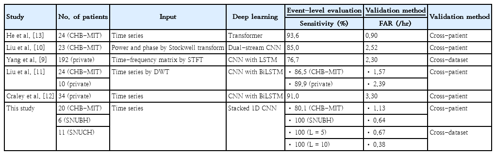
Patient-independent seizure detection performance compared with recent state-of-the-art studies based on deep learning techniques using scalp electroencephalography recordings
Our external validation results are highly encouraging but require cautious interpretation due to the following reasons. Our model employs a strict any-channel trigger logic, where a seizure is flagged if at least one of the 18 channels detects ictal signatures at any time point within the ictal interval. While this design inherently maximizes the model’s sensitivity and minimizes the risk of missing localized seizures, it potentially increases the risk of FAs. In addition, given the relatively small sample size of the external validation cohort, these results may not fully extrapolate to a larger, more heterogeneous clinical population. Lastly, the exclusion of short seizures may have positively influenced the results considering our model’s aforementioned temporal detection mechanism. However, we anticipate that a 30-second duration is sufficiently long to filter out transient motion artifacts (e.g., movement, chewing) and reliably confirm true ictal signals, particularly for wearable seizure detection.
3. Clinical usability for wearable seizure detection
Home-based EEG monitoring with wearable devices for seizure detection has been widely proposed as an alternative diagnostic tool for patients with refractory epilepsy in out-of-hospital settings [24-26]. The use of multiple electrodes has been recognized as one of the most critical challenges in EEG-based wearable seizure detection [7]. Therefore, several recent studies have proposed scalp EEG monitoring using a small number of channels placed behind the ear for wearable seizure detection [27-31]. Although a limited number of channels covers only localized brain regions, previous findings suggest that even a single electrode can detect the majority of electrographic seizures when positioned appropriately [29]. In addition, previous work demonstrated that single-channel seizure detection performance can be comparable to multichannel detection once epileptologists have preconfirmed patients’ seizure onset locations, as shown in a prior patient-specific seizure detection study using the CHB-MIT dataset [21].
To investigate seizure occurrence across entire EEG recordings, the seizure detection model was applied to individual channels in parallel. In focal seizures, EEG recordings from individual channels tend to exhibit distinct electrographic characteristics depending on seizure onset zones and propagation patterns. Therefore, seizures may be more efficiently detected by selecting a limited number of channels that demonstrate clear seizure signatures rather than by using a full-montage configuration. If epileptologists identify high-priority ictal regions in patients with focal epilepsy in advance (e.g., the left parietal and temporal regions shown in Fig. 2 and Supplementary Fig. 5), wearable devices may require only a small number of electrodes corresponding to those regions. The degree of separation between ictal and interictal features in individual channels can quantitatively support appropriate selection of high-priority regions, as illustrated in Fig. 3. Although the number and placement of electrodes may vary among patients, the same pretrained deep learning model can detect seizures across multiple patients by focusing on their ictal regions. In addition, this approach can mitigate situations in which one or several electrodes become unexpectedly detached from the patient’s scalp, because the model continuously monitors seizure occurrence in individual channels in parallel, regardless of electrode detachment, particularly in long-term EEG monitoring with wearable devices.
In the external validation, the effect of detection length on seizure detection performance was examined, demonstrating that doubling the detection length led to a reduction in the FAR of more than 40%, without loss of sensitivity and with only a slight increase in latency. To detect a seizure, the deep learning model must generate ictal probabilities that exceed a predefined threshold continuously for a duration longer than the specified detection length. However, increasing the detection length may cause the model to miss brief ictal characteristics, particularly those occurring near seizure onset, resulting in variability in detection latency for the first seizure of SNUCH_E11 shown in Supplementary Fig. 8. Therefore, detection length can be adjusted as a key parameter in wearable seizure detection systems, especially for patients who are highly sensitive to FAs during EEG monitoring.
4. Limitations and future work
This study has several limitations. First, focal-onset ictal EEG segments were acquired from 2 internal datasets, and external validation was conducted using a single external dataset with a relatively small number of patients. Second, the approach was designed exclusively for the detection of electrographic focal seizures on scalp EEG recordings. Third, the objective was to extract temporal features related to ictal EEG segments within ictal intervals to determine seizure occurrence rather than to precisely localize seizure onset.
In future work, the focal-onset ictal EEG segment dataset will be expanded evenly across various pediatric age groups through multi-institutional collaboration to strengthen the model’s seizure detection capability. We may need to consider the quantitative degree of consensus between 3 or more epileptologists for data quality enhancement. Furthermore, the approach should be evaluated in a larger patient cohort to ensure robust generalizability. However, it may be unrealistic to apply patient-independent seizure detection to all types of epilepsy syndromes. In this study, patients with focal epilepsy exhibiting distinct electrographic seizure signatures on scalp EEG recordings were selected as the primary target population. Therefore, additional external validation will focus on this patient group rather than on patients with excessive movements or markedly suppressed EEG activity. In this process, an understanding of seizure semiology is expected to be highly beneficial for strategizing multimodal sensor combinations in wearable seizure detection.
In conclusion, patient-independent seizure detection achieved a mean sensitivity of 100% with a FAR ≤0.67/hr for 11 patients with focal epilepsy from an external tertiary medical institution using the final seizure detection model trained on 1,604 electrographic ictal EEG segments annotated by epileptologists. The results demonstrate that ictal EEG segments representing diverse seizure patterns play a pivotal role as core reference knowledge for deep learning models in identifying seizure characteristics in individual channels through cross-patient and cross-dataset evaluation strategies. This study represents the first effort to utilize a set of expert-labeled ictal EEG segments as a fundamental reference source for a patient-independent seizure detector to investigate seizure occurrence in scalp EEG recordings. Furthermore, the proposed approach may represent a state-of-the-art patient-independent strategy for selected patients who require continuous seizure monitoring, particularly in wearable seizure detection systems with limited EEG channels.
Supplementary materials
Supplementary methods, Supplementary Tables 1-4, and Supplementary Figs. 1-8 are available at https://doi.org/10.3345/cep.2026.01011.
Patient information of the 23 patients from the CHB-MIT dataset for focal onset ictal EEG segment annotation.
Patient information of the 12 patients from the SNUBH dataset for focal onset ictal EEG segment annotation
Seizure detection performance of the internal validation for the 20 patients from the CHB-MIT dataset
Seizure detection performance of the internal validation for the 6 patients from the SNUBH dataset
Postprocessing procedures in individual channels for event-level evaluation. We select smoothed model outputs equal or higher than a threshold, merge the above-threshold outputs if they are separated each other less than 30 seconds, and select and binarize the outputs if they last longer than a detection length of L (left-side panel). We compute the union of the 18 sets of postprocessed outputs yielding one final set of model-generated ictal intervals for each patient (right-side panel).
A detailed seizure detection model architecture. A 2-second EEG time series is fed into the input layer of the stacked 1-dimensional (1D) CNN architecture as an input signal. Features are extracted through 2 1D CNN modules with different filter sizes in parallel. Two 128-length feature vectors from the 2 modules are concatenated into a final 256-length feature vector feeding into the fully-connected layer to yield an ictal probability from 0 to 1. EEG, electroencephalography; CNN, convolutional neural network.
A representative result of focal onset ictal EEG segment annotation for the first seizure with a length of 67 seconds (the first 38 seconds is shown here) of the chb05 in the CHB-MIT dataset. A greencolored vertical dashed line represents a seizure onset. Yellow-colored boxes represent single-channel-level ictal EEG segments annotated by epileptologists. EEG, electroencephalography; CHB-MIT, Children’s Hospital Boston- Massachusetts Institute of Technology.
A representative result of focal onset ictal EEG segment annotation for the first seizure with a length of 25 seconds of the SNUBH03 in the SNUBH dataset. Green- and red-colored vertical dashed lines represent a seizure onset and termination, respectively. Yellow-colored boxes represent single-channel-level ictal EEG segments annotated by epileptologists. EEG, electroencephalography; SNUBH, Seoul National University Bundang Hospital.
A representative result of a true positive in the event-level evaluation corresponding to the first seizure of the SNUCH_E05 (rescaled time axis of Fig. 2). Blue- and red-colored vertical dashed lines represent epileptologist-annotated (58 seconds) and model-generated (47 seconds) ictal intervals, respectively. (A and B) The upper panels in represent EEG time series with distinct seizure characteristics particularly in the left hemisphere (red-colored channel names and signals). The lower panels represent postprocessed outcomes that blue- and red-colored solid lines represent epileptologist-annotated and channel-wise model-generated ictal intervals, respectively. Gray-colored solid lines represent ictal probabilities for individual channels. SNUCH, Seoul National University Children’s Hospital; EEG, electroencephalography.
Representative false negatives (undetected seizure) missed by our detection model. Panels A and B represent low voltage fast activities evolving into seizure patterns in the chb20. Panels C and D represent rhythmic sharp wave discharges and delta waves with minimal EEG evolution in the chb13. Blue-colored vertical dashed lines represent epileptologist-annotated ictal intervals. EEG, electroencephalography.
Representative false alarms incorrectly identified as seizures by our detection model. Panels A and B represent seizure-like evolution confined to one electrode in the SNUBH09 and chb13, respectively. Panel C represents high voltage muscle artifacts in the chb13. Panel D represents rhythmic chewing artifacts in the SNUBH09. Red-colored vertical dashed lines represent model-generated ictal intervals, along with detected channels and intervals (red-colored channel names and signals). SNUBH, Seoul National University Bundang Hospital.
A representative case of a change of latency in accordance with the detection length (L) in the event-level evaluation corresponding to the first seizure of the SNUCH_E11. In the left-sided figure with L = 5 seconds, green-colored vertical dashed lines represent model-generated ictal intervals with a length of 8 seconds. A red-colored vertical dashed line represents an onset of another model-generated ictal interval next to the green-colored interval. A blue-colored vertical dashed line represents an onset of an epileptologist-annotated ictal interval. In the right-sided figure with L = 10 seconds, the green-colored interval at F4-C4 was not detected by our model because its length was less than L. Detected channels are represented as red-colored channel names. Gray-colored solid lines represent ictal probabilities for individual channels. SNUCH, Seoul National University Children’s Hospital.
Notes
Conflicts of interest
No potential conflict of interest relevant to this article was reported.
Funding
This work was supported by the New Faculty Startup Fund from Seoul National University under Grant 800-20240583.
Author contribution
Conceptualization: HK; Data curation: JC, AC, HK, BCL; Formal analysis: YGC; Funding acquisition: HK; Methodology: YGC, JC, HK; Project administration: HK; Writing - original draft: YGC, JC, HK; Writing - review & editing: YGC, JC, AC, HK, BCL