Red cell distribution width and reticulocyte hemoglobin content as predictors of iron depletion in children with cyanotic congenital heart disease
Article information

Abstract
Background
Iron-deficiency anemia is challenging to diagnose in patients with cyanotic congenital heart disease (CCHD) because of the high hemoglobin concentration as a compensatory mechanism for cyanosis.
Purpose
This study aimed to identify predictive parameters of iron depletion status and assess the prevalence of iron deficiency in patients with CCHD.
Methods
A descriptive study enrolled patients with CCHD aged 6 months to 15 years between September 2022 and September 2023. Participants were categorized into iron depletion and iron sufficiency groups defined by a serum ferritin level <30 ng/mL or transferrin saturation <15%. The clinical characteristics and laboratory parameters were compared between groups. The Youden index was used to determine the optimal cutoff points, maximizing both sensitivity and specificity for the red cell indices.
Results
Among the 110 enrolled patients with CCHD, 28 (25.5%) had iron depletion. While hemoglobin (Hb) and hematocrit (Hct) levels were comparable between groups, the iron-depleted group exhibited significantly lower reticulocyte hemoglobin content (CHr) and higher red cell distribution width (RDW). A multivariate analysis identified an RDW ≥18.8% (odds ratio [OR], 7.79; P=0.001) and CHr <28 pg (OR, 4.97; P=0.004) as independent predictors of iron depletion. The diagnostic accuracy improved to 85.4% when these parameters were combined.
Conclusion
Iron depletion is highly prevalent in patients with CCHD. Because conventional parameters, such as Hb and Hct, fail to differentiate iron status owing to compensatory mechanisms, CHr and RDW serveas effective predictive tools. This combination offers a practical diagnostic alternative, especially in resource-limited settings, where standard iron studies are unavailable.
Key message
Question: Are red cell distribution width (RDW) and reticulocyte hemoglobin content (CHr) reliable alternative markers for detecting iron depletion in children with cyanotic congenital heart disease, whose status is often masked by compensatory polycythemia?
Finding: Multivariate analysis demonstrated that RDW (≥18.8%) and CHr (<28 pg) are strong independent predictors of iron depletion in this vulnerable population.
Meaning: Combined use of these accessible markers facilitates earlier diagnosis and improves clinical management in resource-limited healthcare facilities.
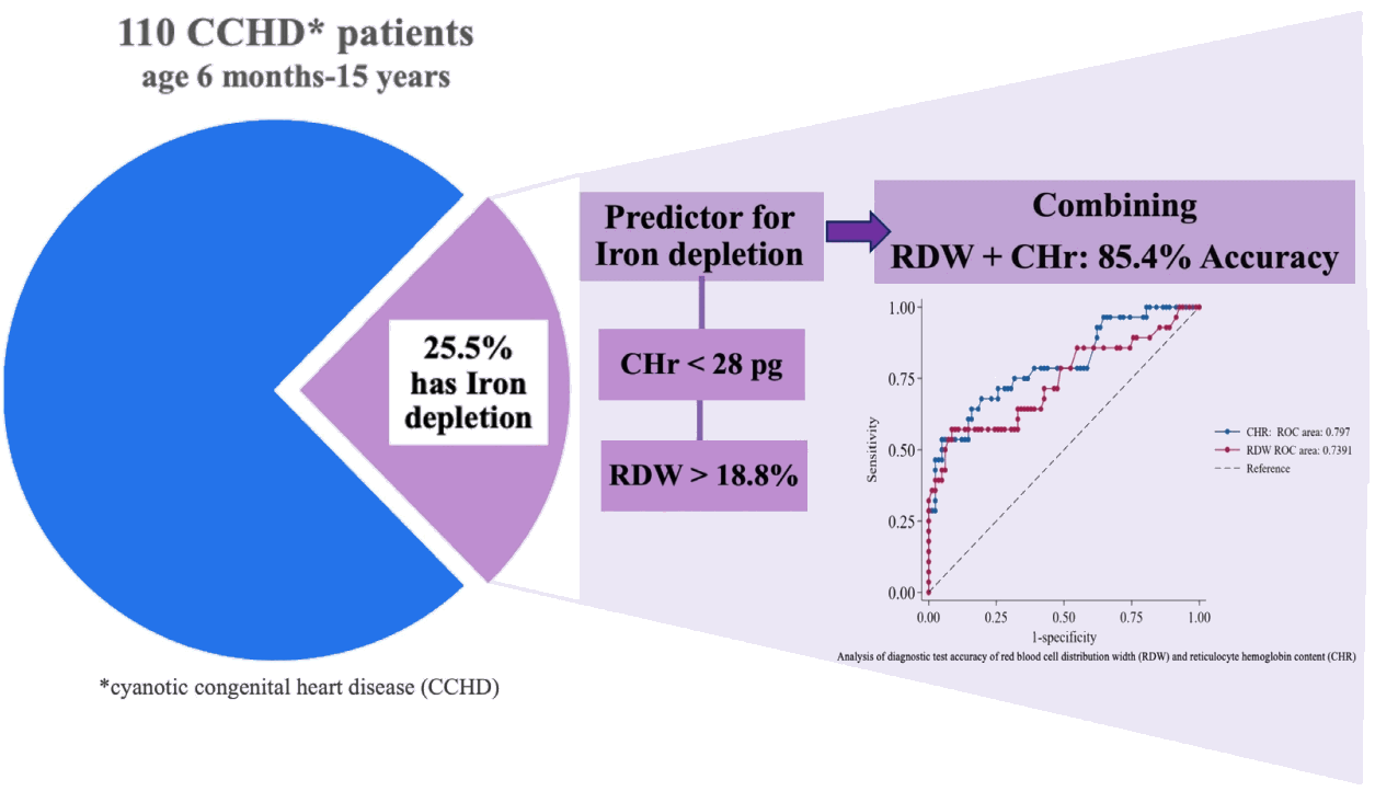
Graphical abstract. CHr, reticulocyte hemoglobin content; RDW, red blood cell distribution width.
Introduction
Iron is one of the essential nutrients that is crucial for growth and development during childhood. Insufficient iron levels result in anemia, leading to prolonged delays in brain development and hindering overall growth [1]. In cyanotic congenital heart disease (CCHD) patients, in addition to impairment of development, iron insufficiency is associated with a higher rate of hypercyanotic spell [2]. According to 2019 data from the World Health Organization, the global prevalence of iron-deficiency anemia in children aged 6 months to 5 years is 39.8%, with a rate of 24.9% reported in Thailand [3].
The diagnosis of anemia is established by measuring hematocrit levels that fall below the 5th percentile within the same age group of the population [4]. Individuals with CCHD tend to exhibit higher hemoglobin concentrations than the general population due to a compensatory mechanism for baseline cyanosis to ensure sufficient oxygen delivery [5,6]. In children with CCHD, there are no specific cutoff points for hematocrit levels for diagnosing anemia. Utilizing red blood cell (RBC) count, hemoglobin, and hematocrit levels to assist in diagnosing iron-deficiency status would present challenges.
Previously, the gold standard for diagnosing iron deficiency involved bone marrow aspiration [7,8]. However, due to invasive nature and difficulty of the procedure, the serum iron (SI) measurement was developed and has since become the standard tool for diagnosing iron deficiency [9,10]. In routine clinical practice, serum ferritin (SF), SI, and total iron-binding capacity (TIBC) are commonly used to diagnose iron deficiency [1,8]. Due to cost-effectiveness and availability especially in limited resources setting, complete blood count (CBC) is often used to add to the diagnosis of iron-deficiency anemia [9,10]. Rather than SF, SI, and TIBC, reticulocyte hemoglobin content (CHr) is accepted as another precise and widely-available parameter for accessing iron status, which is less affected by the inflammation process than SF [11,12].
This study aims to determine the prevalence of iron depletion in CCHD patients and identify specific red cell indices that can serve as diagnostic tools for detecting iron deficiency in individuals with cyanotic heart conditions.
Methods
We conducted a descriptive study, collecting data from patients at King Chulalongkorn Memorial Hospital from September 2022 to September 2023. The inclusion criteria were patients aged between 6 months and 15 years with a confirmed diagnosis of CCHD. The CCHD was defined as presence of cardiac structural malformation since birth, confirmed with echocardiography by pediatric cardiologist, and oxygen saturation (SpO2) below 95% in room air.
The exclusion criteria involved participants who had received blood transfusion within the last 6 months, prior diagnosed with thalassemia or iron-deficiency anemia, and potential iron malabsorption conditions (such as previous history of bowel resection).
The minimum sample size was determined using Cochran formula. Based on an estimated prevalence of 30% and a 10% margin of error at a 95% confidence level (CI), a minimum of 81 participants was required for this study.
In our study, we collected the demographic data, including sex, age, weight, height, underlying conditions, SpO2 in room air, previous history of iron supplementation, and, if applicable, details regarding the dose and compliance of iron supplements. Blood samples were obtained to conduct a comprehensive analysis, which included a CBC, reticulocyte count (incorporating CHr, SI, SF, and TIBC). Subsequently, patients were categorized into 2 groups: iron depletion and noniron depletion. The iron depletion group consisted of participants with SF levels below 30 ng/mL or transferrin saturation below 15%. The characteristic data and laboratory results of these 2 groups were compared. Wilcoxon rank-sum test was used to compare continuous data, and chi-square or Fisher exact test were used to compare categorical data.
Youden index method was performed to determine the optimal cutoff points with the best sensitivity and specificity for biomarkers. The performance of biomarkers was evaluated by the receiver operating characteristic curve. Additionally, a multivariate logistic regression method was utilized to identify significant parameters serving as predictors for iron depletion status. Covariates with a P value <0.1 in the univariate analysis were entered into the multivariate model using a backward stepwise selection method.
This study was approved by the institutional review board (IRB) of Faculty of Medicine, Chulalongkorn University (IRB No. 0407/65).
Results
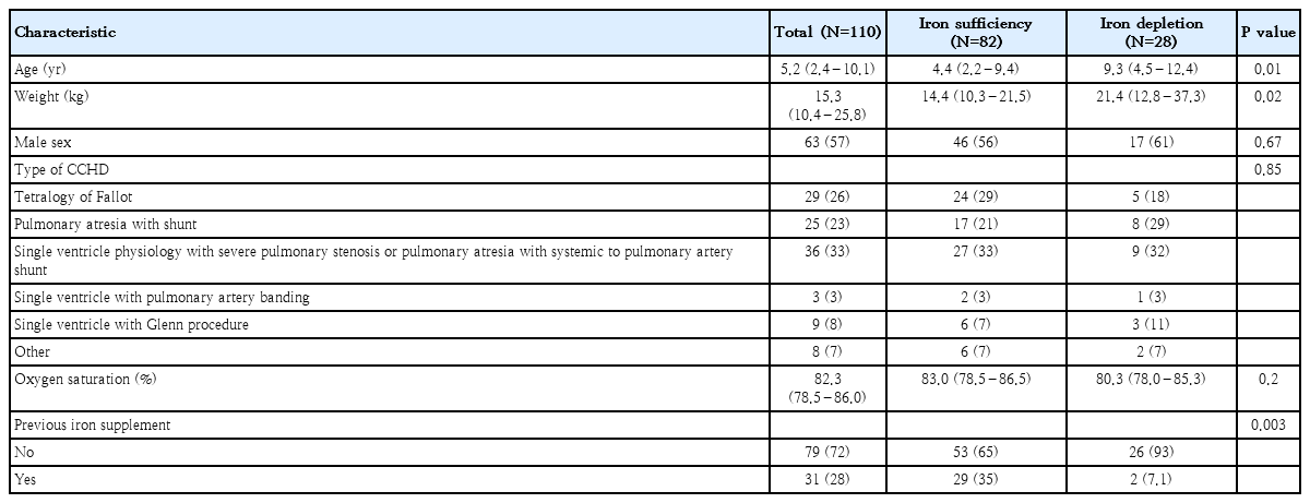
One hundred and 10 participants were recruited for this study. The demographic details were summarized in Table 1. The mean age of participants was 5.2 years (range, 2.4–10.1 years), predominantly male (57.3%). The most common type of CCHD is single ventricle physiology with severe pulmonary stenosis or pulmonary atresia with systemic to pulmonary artery shunt, accounting for 33%, followed by tetralogy of Fallot. Thirty-one patients (28.2%) among all participants received iron supplements in the past 6 months.
Baseline characteristics of participants in the iron depletion and iron sufficiency groups
The results indicated a prevalence of iron depletion at 25.5% among CCHD patients. The median age and weight are higher in iron depletion group, but there is no significant difference in SpO2 between both groups. Notably, the prevalence of iron depletion was significantly lower in patients who had previously taken iron supplements with an average dose of 2.65 mg/kg/day.
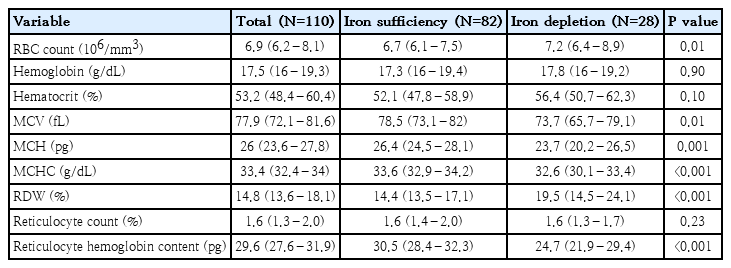
The laboratory analysis included red cell indices, reticulocyte count, and CHr were demonstrated in Table 2. Although there were no significant differences in hemoglobin, hematocrit and percentage of reticulocyte count between both group, specific RBC indices showed distinctions.
Red blood cells indexes in children with cyanotic congenital heart disease by study group
The RBC count was higher in the iron depletion group (P=0.01), and similarly, the red cell distribution width (RDW) was elevated in the iron depletion group (P<0.001). Additionally, the mean corpuscular volume (MCV), mean corpuscular hemoglobin (MCH), and mean corpuscular hemoglobin concentration (MCHC) were significantly lower in the iron depletion group (P=0.01, P<0.001, and P<0.001, respectively). CHr was also significantly lower in the iron depletion group.
Based on the data presented in Table 2, Youden index method was performed to assess the effectiveness of various biomarkers in predicting iron depletion status within our study population. The results shown in Table 3 indicated that MCH, RDW, and CHr exhibit good specificity for diagnosing iron depletion. Specifically, RDW more than 18.8% and CHr below 28 pg demonstrated good performance as predictors for identifying iron depletion status, indicated by an area under the curve greater than 0.7. These findings suggested that RDW and CHr might be reliable indicators for predicting iron depletion.
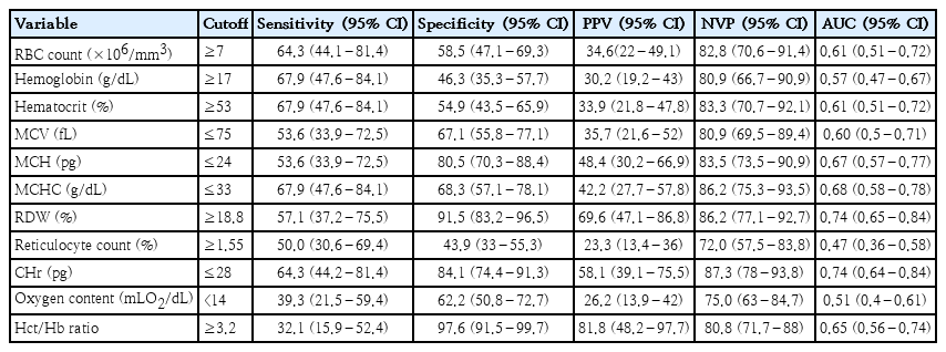
Performance of biomarkers for prediction iron depletion status in children with cyanotic congenital heart disease
Subsequently, univariate and multivariate logistic regression were carried out to identify parameters significantly correlated with the iron depletion status of the patients. As indicated in Table 4 and Fig. 1, the results reveal that RDW of more than 18.8% (odds ratio [OR], 7.79; 95% CI, 2.46–25.13; P=0.001) and CHr of less than 28 pg (OR, 4.97; 95% CI, 1.67–14.84; P=0.004) were significantly associated with iron depletion.
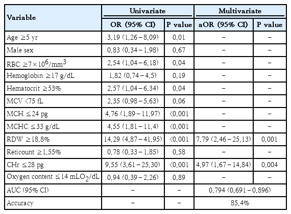
Predictors of iron depletion status
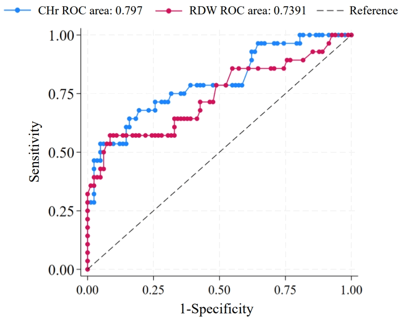
Analysis of diagnostic test accuracy of red blood cell distribution width and reticulocyte hemoglobin content. CHr, reticulocyte hemoglobin content; ROC, receiver operating characteristic; RDW, red blood cell distribution width.
Remarkably, the effectiveness of diagnosing iron depletion was enhanced with the accuracy of 85.4% when combining RDW and CHr as predictors.
Discussion
In this study, the prevalence of iron depletion among patients with CCHD was 25.5%, which is comparable to the reported prevalence of iron-deficiency anemia in healthy Thai children [3], regardless of prior iron supplementation history. Notably, when specifically analyzing the subgroup without a history of iron supplementation, the prevalence increased to 32.9%. Across various studies of CCHD populations, reported prevalence rates of iron-deficiency anemia vary widely, ranging from 19.7% to 46% [13-16]. This significant discrepancy is primarily attributed to the lack of a standardized diagnostic definition, as prevalence figures remain highly dependent on the specific biochemical criteria and cutoff points employed by different investigators. We demonstrated that hemoglobin and hematocrit levels were not significantly associated with iron depletion status in cyanotic heart patients. This corresponds with our hypothesis of an increase in hemoglobin and hematocrit as a physiological response to cyanosis [5,6,17], and may complicate the interpretation of iron-deficiency anemia in CCHD patients. However, many other RBC parameters including low MCV, low MCH, low MCHC, high RDW, and high CHr, in the iron depletion group were consistent with findings from other studies [2,18-23]. This supports the reliability of the parameters as indicators for iron depletion. In Thailand, many hospitals lack the capacity to conduct iron studies and thus resort to outsourcing them. Moreover, from an economic perspective, within our medical center, the cost of iron studies is approximately 4 to 5 times that of a CBC plus a reticulocyte count. Selecting cases with a high probability of iron deficiency based on CBC parameters and reticulocyte count would be beneficial. In this study, we aimed to identify predicting factors for iron depletion from RBC parameters. Multivariate analysis confirmed that CHr and RDW are robust independent predictors of iron depletion in our cohort. Although RDW can be elevated in various conditions, such as hemolysis, hemoglobinopathies, vitamin B12 or folate deficiencies, and heart failure [24], our findings align with those of Bandyopadhyay et al. [14], who reported RDW as a reliable indicator for iron deficiency in CCHD. Furthermore, our results complement the work of Cheng et al. [16], who reported that reticulocyte hemoglobin equivalent could be utilized as an iron status parameter in pediatric cyanotic heart disease, with cutoff values of 28.8 pg for iron deficiency. In this study, a CHr ≤28 pg and an RDW ≥18.8% were significantly associated with iron depletion. Notably, the diagnostic performance was further enhanced when these parameters were used in combination, suggesting that an integrated approach provides superior accuracy in detecting iron deficiency within this complex patient population. Although, the previous studies have indicated that MCV is lower in iron depletion patients [18,21]. Our data did not demonstrate good specificity of MCV to iron depletion, possibly influenced by the high prevalence of thalassemia in Thailand [25,26].
Interestingly, the RBC count was significantly higher in the iron depletion group. One proposed hypothesis to explain this observation is the overproduction of RBCs secondary to chronic hypoxemia [5,6]. However, insufficient iron may lead to an increase in microcytic RBCs, resulting in low hemoglobin content, while also raising the overall RBC count [13].
Notably, 28.2% of the subjects in this study had previously received iron supplementation at an average dose of 2.65 mg/kg/day. This nontherapeutic supplementation dose [9] is a common practice in our center to provide adequate substrate for proper RBC production, aiming to compensate for hypoxia in cyanotic heart disease (CCHD) patients. One potential limitation of our study is that iron supplementation may interfere with laboratory results. To address this potential confounder, we conducted a subgroup analysis, focusing on patients who did not receive iron supplementation. The findings from multivariate logistic regression still indicated that RDW and CHr were significantly different. This strengthens our initial conclusions, suggesting that RDW and CHr are reliable indicators for predicting iron depletion status in CCHD patients, even when considering the influence of iron supplementation. We also observed a significantly low prevalence of iron depletion in the group of patients who received iron supplementation (P<0.003). Among the 31 patients who received iron supplement doses, 2 patients who still exhibited iron depletion were found to have poor compliance with the medication. Additionally, none of the 31 patients who had taken iron supplements reported serious side effects such as gastrointestinal infection [27] that interfered with their daily lives. This information suggests the potential benefits of iron supplementation in preventing iron depletion and indicates a generally well-tolerated profile of iron supplements among the studied patients. For clinical application, in settings where iron studies or laboratory investigations cannot be performed, dispensing iron supplement doses to CCHD patients may be considered.
In conclusion, the result from this research suggests that using CHr at a cut-point ≤28 pg and RDW ≥18.8% can be utilized to predict iron depletion status. The relation to iron depletion status is notably stronger when these parameters are used in combination.
Notes
Conflicts of interest
No potential conflict of interest relevant to this article was reported.
Funding
All the phases of this study were supported by the Ratchadaphiseksomphot Endowment Fund (MDCU GA66/09).
Author contribution
Conceptualization: HP, KC, KW; Data curation: HP, KW; Formal analysis: HP; Funding acquisition: KW; Methodology: HP, KC, KW; Project administration: HP, KW; Visualization: HP, KW; Writing - original draft: HP, KC, KW; Writing - review & editing: KC, KW, NP, VL, PL