Current review of pediatric Fas-associated death domain protein deficiency: expanding clinical and therapeutic perspectives
Article information

Abstract
Fas-associated death domain protein (FADD) deficiency is a rare inborn error of immunity characterized by dysregulated T-cell proliferation. The clinical spectrum and management of FADD deficiency in children remain incompletely described. This review aimed to synthesize patient-level observational evidence on clinical manifestations, immunological and genetic findings, and treatment outcomes of pediatric patients with FADD deficiency. A literature search was conducted of PubMed, the Cochrane Library, and Scopus from database inception to July 2, 2026, and reference lists of eligible articles were reviewed. Inclusion criterion was articles on human clinical studies of patients with FADD gene mutation. Eligibility screening and data extraction were performed independently. Ten articles describing 18 patients were included. The reported ancestries included South Asian (n=9), European (n=6), and East/Central Asian (n=1). Parental consanguinity was reported in 10 of 12 patients (83.3%). Median age at onset was 0.8 years. Major presentations were fever-related encephalopathy, lymphoproliferation, and invasive pneumococcal disease. Common features included liver dysfunction, seizures, and functional hyposplenism. Regarding immunophenotyping, double-negative T cells were elevated in 9 of 10 patients (90%). Elevated soluble FAS ligand and interleukin-10 levels and defective lymphocyte apoptosis were observed in all tested patients; most patients had elevated vitamin B12 levels. The most common pathogenic variant was c.350G>A, followed by c.315T>G. Six patients died (33%) at a median age of 0.8 years. Mortality was high among patients with invasive pneumococcal disease; no deaths were reported among those with a lymphoproliferative phenotype or among the 2 hematopoietic stem cell transplantation recipients. FADD deficiency ranges from early-onset fever-related encephalopathy and fulminant sepsis to lymphoproliferative phenotypes. This review emphasizes the importance of recognizing FADD deficiency in children presenting with recurrent febrile encephalopathy, liver dysfunction, and a history of consanguinity and highlights prompt genetic evaluation and hematopoietic stem cell transplantation as potential curative therapies.
Key message
Fas-associated death domain protein deficiency expands beyond a severe infantile immunodeficiency to a broader clinical spectrum in children involving autoimmunity, autoinflammation, and susceptibility to bacterial and viral infections. This review highlights recurrent febrile encephalopathy, invasive pneumococcal disease, and liver dysfunction as key features and identifies later-onset and self-limiting phenotypes.
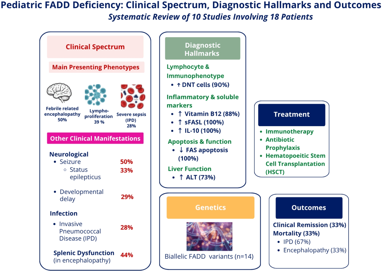
Graphical abstract. DNT, double-negative T cell; FAS, first apoptosis signal; sFASL, soluble FAS ligand; IL, interleukin; ALT, alanine aminotransferase; FADD, Fas-associated death domain protein.
Introduction
An intact Fas-associated death domain (FADD) pathway is crucial for a balanced immune regulation and effective immune response against pathogens. This is achieved by the induction of apoptosis of self-reactive T cells that prevents the proliferation of autoreactive T cells. FADD, caspase-8, and RIPK1 (receptor-interacting serine/threonine protein kinase 1) are components of the FADDosome complex. When induced by tumor necrosis factor-related apoptosis-inducing ligand (TRAIL) and FAS ligand (FASL), the FADD pathway promotes apoptosis and inflammatory cytokine production via the nuclear factor-kappa B pathway [1]. It also plays an important role in immunity against viruses via positive feedback provided by type 1 interferon signaling and hematopoiesis [2,3]. The FADD protein is expressed in a wide variety of tissues, including blood cells and the brain, heart, liver, spleen, and lymph nodes. FADD deficiency was first described in 2010, when 4 children from a consanguineous family presented with autoimmune lymphoproliferative syndrome (ALPS)-like features along with congenital cardiac malformation, functional hyposplenism, encephalopathy, hepatic dysfunction, and recurrent bacterial and viral infections [2].
Since the first identification of FADD deficiency almost 15 years ago, there have been only sporadic reports of this rare autosomal recessive condition. Although the role of the FADD pathway in T-cell apoptosis and immune regulation is well-documented, the clinical spectrum and treatment outcomes of FADD deficiency remain poorly understood. A 2022 review focused on a small cohort of patients and provided incomplete characterization of immunological features and therapeutic responses, particularly for hematopoietic stem cell transplantation (HSCT) [4].
Here we aimed to review published cases in the literature, summarize their clinical features and laboratory findings, and highlight current treatments for FADD deficiency in children, including potential curative approaches such as HSCT. This study aimed to raise awareness of this syndromic inborn error of immunity (IEI) and explore potential options for curative therapy such as HSCT.
Methods
1. Eligibility criteria
This study represents an updated review of the published literature. Articles were included if they involved human clinical studies of patients with biallelic mutations of the FADD gene. All original human clinical and nonexperimental descriptive studies, including cohort studies, case series, case reports, and clinical trials, were included. Studies involving non-FADD mutations and nonclinical in vitro studies were excluded.
2. Search sources
This literature search spanned multiple databases, including PubMed, the Cochrane Library, and Scopus. The search was conducted from database inception to July 2, 2026. The reference lists of the included articles were screened for additional studies. No filters were applied or restrictions placed on publication dates or language to ensure comprehensive search coverage. The full search strategies used for each database are provided in the Supplementary Material.
3. Selection process and data collection
All of the retrieved citations were compiled using the EndNote 20 citation manager (Clarivate, USA). Duplicate entries were identified and removed using both EndNote automated tools and manual review.
The selection process was conducted independently by 2 reviewers in 2 stages:
(1) Title and abstract screening: Articles were reviewed for eligibility based on relevance. To facilitate the study selection process, the eligibility screening was performed using Rayyan, a web-based systematic review platform.
(2) Full-text review: Articles passing the initial screen underwent a thorough evaluation. A total of 1,194 records were initially identified through database searches (n=1,000) and the reference lists of the relevant articles (n=194). After the exclusion of 334 duplicate records and 841 records that did not meet the eligibility criteria, 19 reports were retrieved and subjected to full-text assessment. The full-text review process excluded 9 articles; thus, 10 studies were subjected to the final review. The article inclusion and exclusion process is shown in the PRISMA (Preferred Reporting Items for Systematic Reviews and Meta-analyses) flowchart (Fig. 1).
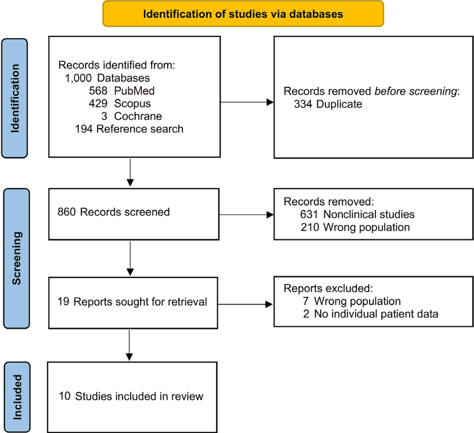
PRISMA (Preferred Reporting Items for Systematic Reviews and Metaanalyses) flowchart illustrating the article screening process.
4. Data extraction and management
Individual patient-level data were extracted from the full-text articles and supplementary materials, and all data were verified by multiple reviewers for accuracy. The extracted data variables included participant demographics, clinical features, laboratory findings, genetic findings, interventions, and outcomes.
Duplicate cases in the selected articles were identified by cross-referencing of the demographic characteristics, clinical features, family pedigree, and genotype. The duplicate cases were P8 (Kohn III.5, Meer, Vogel) [4-6], P16 (Pelle P2, Rensing-Ehl P15), P17 (Pellé P3, Rensing-Ehl P16), and P18 (Pelle P4, Rensing-Ehl P10) [7,8]. These duplicate reports were consolidated and each patient was counted only once in the cohort. The corresponding authors of the reviewed articles were contacted to clarify any missing data and address any uncertainties. Any unresolved missing information in the dataset was handled using pairwise deletion during the analysis.
5. Statistical methods
This study was designed as a systematic review with a descriptive pooled analysis of individual patient data. Continuous variables are reported as median and interquartile range (IQR), while categorical variables are summarized as frequencies and percentages. All descriptive statistics were analyzed using IBM SPSS Statistics ver. 26.0 (IBM Co., USA).
6. Major updates in this review
This updated review expands the prior review by Meer et al. [4] in 2022, which analyzed only 4 patients. In contrast, the current review provided a comprehensive analysis of 18 patients, including 10 probands and their affected siblings. This review thoroughly describes patient demographics, clinical features, neuroimaging findings, biochemical and immunological parameters, genetic findings, interventions, and treatment outcomes and offers a more detailed understanding of FADD deficiency.
Results
1. Patient characteristics and genetic information
The cohort included 18 patients from 10 families, with a slight female predominance (56%). The median age at disease onset was 0.8 years. Most patients were from South Asia (9 of 16 [56%]) or Europe (6 of 16 [38%]). A high proportion (10 of 12 [83%]) of the patients were from consanguineous families. Additional demographic details are shown in Table 1.
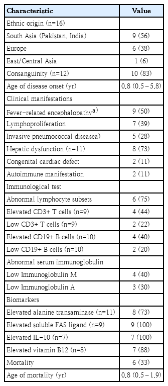
Demographics and clinical characteristics of patients with FADD deficiency
Genetic diagnoses were confirmed in 14 patients (78%). Four patients from 3 families with confirmed FADD mutations did not undergo genetic testing because they died before the proband in each family was diagnosed. Their clinical phenotypes were consistent with FADD deficiency. Therefore, they were inferred to carry the same familial mutation as their respective probands. Whole exome sequencing (WES) was the most frequently used diagnostic test, performed in 71% (10 of 14) of the patients, followed by Sanger sequencing in 64% (9 of 14).
Among the patients with confirmed FADD mutations (n=14), homozygous mutations were identified in 9 patients (64%), 4 cases (29%) of heterozygous mutations, and 1 (7%) compound heterozygous mutation. A germline heterozygous FADD variant with somatic loss of heterozygosity via uniparental disomy was identified in 4 patients (4 of 14 [29%]), all of whom had lymphoproliferation [7]. This mutation was identified using array-based comparative genomic hybridization of DNA from sorted double-negative T cells. All mutations were localized to exon 2 of the FADD gene, and the most common variant was c.350G>A (p.R117H), which was identified in 43% (6 of 14) of patients, followed by c.315T>G (p.C105W) in 29% (4 of 14). Table 2 summarizes the frequencies of FADD mutations reported in this review.
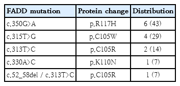
Distribution of reported FADD pathogenic variants
2. Clinical manifestations
Recurrent fever-related encephalopathy was reported in 50% of the patients (9 of 18); all experienced altered consciousness and seizures following febrile episodes. Among the patients with febrile-related encephalopathy (n=9), 3 (33%) developed status epilepticus. Febrile-related encephalopathy was triggered by specific events, for example, acute viral infections, occurring in 56% (5 of 9) of cases, or after the measles, mumps, and rubella vaccination (2 of 9 [22%]). The Herpesviridae family accounts for most viral triggers, including varicella-zoster virus, human herpes virus 6, cytomegalovirus (CMV), and Epstein-Barr virus (EBV) [2,5]. Other implicated pathogens include parainfluenza virus and astrovirus. Invasive pneumococcal disease (IPD) was reported in 28% (5 of 18) of the patients. The manifestations included meningitis in 4 patients and sepsis in 1 patient, highlighting the susceptibility of these individuals to severe IPD. Other reported infections included CMV and bacillus Calmette-Guérin (BCG)-itis in 1 patient each. The reported BCG-itis patient, who was less than 1 year of age, presented with lymphoproliferation and subsequently developed BCG-itis following the BCG vaccination [9]. The patient harbored a homozygous FADD c.350G>A (p.Arg117His) pathogenic variant.
Additional findings included lymphoproliferation in 39% (7 of 18) of patients. Autoimmune manifestations are rare in FADD deficiency and were identified in 2 patients who presented with Evans syndrome and autoimmune neutropenia [7,10]. Malignancy was reported in 2 of the 18 patients (11%), comprising EBV-driven diffuse large B-cell lymphoma with disease onset at 36 years of age and stage IV Hodgkin lymphoma with onset at 3 years of age. Both patients were female and showed lymphoproliferation before malignancy development.
Ocular manifestations were reported in only 1 patient in the review cohort [4]. As the disease progressed, the patient experienced visual loss at 7 years of age, with a visual acuity of 20/400 in the right eye and 20/60 in the left eye. Detailed ocular assessments, including fundus photography, fluorescein angiography, and optical coherence tomography, revealed bilateral cystoid macular edema, peripheral retinal ischemia, and neovascularization of the optic disc with tractional retinal detachment and vitreous hemorrhage in the right eye. Congenital heart disease was reported in 11% (2 of 18) of patients, including left-sided superior vena cava draining into the left atrium and pulmonary atresia with a ventricular septal defect [2].
All patients carrying the c.350G>A mutation presented with lymphoproliferation, whereas those with fever-related encephalopathy carried the c.313T>C or c.315T>G mutation. The clinical manifestations of FADD observed in the reviewed cohort are shown in Fig. 2.
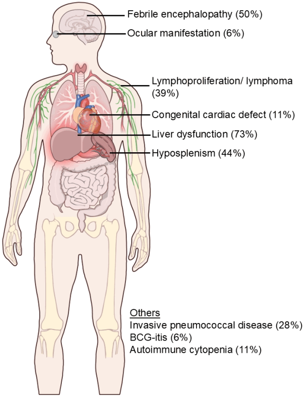
Clinical manifestations of FADD deficiency illustrating the key clinical features observed in patients with FADD deficiency along with their respective frequencies within the reviewed cohort. FADD, Fas-associated death domain; BCG, bacillus Calmette- Guérin. Illustration courtesy of NIAID (https://bioart.niaid.nih.gov/bioart/519).
3. Immunological features
Peripheral lymphocytosis was noted in 63% (5 of 8) of patients, whereas some patients showed abnormal lymphocyte subset proportions, with increased CD3+ T cell (4 of 9 [44%]) or increased CD19+ B-cell (4 of 10 [40%]) counts. Among the patients for whom immunoglobulin data were available, 40% (4 of 10) showed low immunoglobulin M levels and 30% (3 of 10) had low immunoglobulin A levels.
Functional hyposplenism, as evidenced by the presence of Howell-Jolly bodies, was identified in 44% of patients (4 of 9). Functional immune defects included suboptimal antibody production against vaccines in 40% of the tested patients (2 of 5), while all assessed patients demonstrated lymphocyte apoptosis defects with an increased lymphocyte survival rate (n=11; median, 74%; IQR, 50%–92%). Double-negative T-cell (CD4-CD8-TCRαβ) counts were increased in 90% of patients (9 of 10), with a median level of 12% (IQR, 4%–20%).
4. Liver function and biochemical analyses
Liver dysfunction, indicated by elevated transaminase levels, was observed in 73% (8 of 11) of patients. Among those for whom alanine aminotransferase levels were available (n=11), the median was 61 U/L (IQR, 40–331 U/L). Additional biomarker abnormalities included elevated soluble FASL levels (n=9; median, 1,609 pg/mL; IQR, 1,103–3,110 pg/mL), elevated interleukin-10 levels (n=7; median, 302 pg/mL; IQR, 93–431 pg/mL), and elevated vitamin B12 levels in 88% (7 of 8) of cases (median, 2,000 pg/mL; IQR, 1,306–3,748 pg/mL).
5. Neurological features
Neurodevelopmental delay or cognitive dysfunction was reported for 29% (4 of 14) of patients. Abnormal neuroimaging findings were observed in 75% (6 of 8) of the patients. Among them, 38% (3 of 8) exhibited cerebral atrophy, while another 38% (3 of 8) showed white-matter abnormalities, including restricted diffusion or hyperintensities primarily affecting the corpus callosum, corona radiata, or temporo-occipital region.
Serial brain magnetic resonance imaging (MRI) findings were documented for only one patient [11]. Serial brain MRI imaging revealed evolving changes corresponding to recurrent episodes of fever-related encephalopathy. At 12 and 15 years of age, following the first and second encephalopathic episodes, respectively, brain MRI findings were unremarkable and showed no abnormalities. However, at 17 years of age, following the third fever-related encephalopathy episode, brain imaging revealed abnormalities including diffuse T2/fluid-attenuated inversion recovery hyperintensities in the right mesial temporal, occipital, and insular cortices as well as hyperintensities in the left temporo-occipital region. The MRI perfusion sequence revealed marked hyperperfusion suggestive of increased cerebral blood flow. By the time the patient was 20 years old, a follow-up MRI showed right hippocampal head atrophy on T2/fluid-attenuated inversion recovery sequences. These MRI findings suggested that progressive structural brain damage is associated with recurrent encephalopathy episodes.
6. Treatment and clinical outcomes
The management strategies varied across all reported cases. The acute treatment of encephalopathy with 5 days of pulse intravenous methylprednisolone led to disease remission in 2 patients [6,11]. Chronic treatment regimens included oral prednisolone (3 patients), mycophenolate mofetil (MMF; 2 patients), sirolimus (2 patients), and monoclonal antibodies (1 patient). Natalizumab (5 mg/kg) was administered to a patient who developed hemiparesis, dysarthria, and ataxia after an episode of viral-related encephalopathy [6]. After 3 months of treatment, marked clinical improvement was observed. By 1 year, the patient was able to perform all activities of daily living and walk independently and showed no signs of weakness or encephalopathy. However, the dysarthria and ataxia persisted. Despite these residual symptoms, the patient experienced no new neurological complications associated with the viral infections during therapy. Combination therapy consisting of oral prednisolone (2 mg/kg/day) and MMF (1,200 mg/m2/day) was administered to a patient with lymphoproliferative disease to treat ALPS [10]. This treatment resulted in regression of the lymphadenopathy and cessation of the systemic steroids. However, dependency on MMF was observed as lymphocytosis and thrombocytopenia recurred upon its withdrawal. Two patients with ALPS-like lymphoproliferative phenotypes were treated with sirolimus and achieved clinical remission [7]. The reviewed articles did not specify the duration of therapy for patients who received long-term immunomodulators. Prophylactic antibiotics were used in 4 patients, effectively preventing IPD [2]; however, the specific antibiotics used were not reported. Clinical remission was reported in 6 of 18 patients (33%). Of them, 4 achieved posttreatment remission. Among those patients who achieved remission, 4 had an ALPS-like lymphoproliferative phenotype and 2 had febrile-related encephalopathy. Remarkably, 11% of patients (2 of 18) achieved disease resolution (no recurrence of fever-related encephalopathy or lymphoproliferation) without specific treatment.
The overall mortality rate was high: 33% (6 of 18) of patients succumbed to the disease, typically within the first year of life (median age at mortality, 0.8 years; IQR, 0.5–1.9 years). Notably, IPD was the cause of death in 4 of 6 deceased patients (67%). None of the patients who succumbed to the disease received immunotherapy.
7. Hematopoietic stem cell transplantation
HSCT was performed in 2 patients with FADD deficiency [12], both of whom remained free of IPD and febrile encephalopathy posttransplantation.
The first patient, reported by Savic et al. [12], underwent HSCT at 2 years of age using a haploidentical donor (grandfather). Engraftment was performed on day +14. However, incomplete chimerism necessitated a stem cell top-up at 10 months post-HSCT, which was performed as an unconditioned unmanipulated boost [13]. Despite this intervention, the patient achieved only low-level mixed chimerism after the second procedure. A chimerism analysis after the stem cell top-up showed donor T cell, B cell, and myeloid chimerism at 29%, 5%, and 12%, respectively. Posttransplant complications included vasculopathy of the small muscular arteries by day +330, and the patient presented with gastrointestinal bleeding and abdominal pain. The patient then required a bowel resection and sirolimus treatment and was dependent on parenteral nutrition [12]. A histopathological examination of the resected bowel tissue confirmed this diagnosis.
The second patient reported by Savic et al. [12] underwent HSCT using a matched sibling donor. Engraftment was achieved by day +25 and donor chimerism reached 100% by day +28. Grade II skin graft-versus-host disease (GVHD) developed on day +28 but resolved by day +42. However, the patient exhibited persistent leukodystrophy on MRI indicative of ongoing neurological challenges [12]. Table 3 presents the details of the preconditioning chemotherapy regimen, engraftment, and donor chimerism. GVHD prophylaxis was not discussed in any of the reviewed studies.
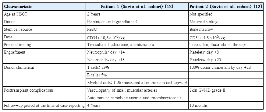
Hematopoietic stem cell transplantation details and outcomes of FADD deficiency
Discussion
This updated review provides a comprehensive analysis of 18 pediatric patients with FADD deficiency, a rare autosomal recessive immunological disorder. This review expands on the previous literature by including 10 probands and their affected siblings. FADD deficiency should be suspected in patients of Asian origin, particularly in the presence of consanguinity and symptom onset during infancy. Its key clinical manifestations include recurrent fever-related encephalopathy, recurrent IPD infections, and hepatic dysfunction. A family history of similar presentations further supported the likelihood of this diagnosis.
In suspected cases, laboratory evaluations should include a complete blood count to assess lymphocytosis and a peripheral blood smear to identify Howell-Jolly bodies, which may indicate functional hyposplenism. Liver function tests are also recommended because elevated transaminase levels may reflect hepatic dysfunction associated with FADD deficiency. Immunological assessments should include flow cytometry for lymphocyte immunophenotyping, with particular attention paid to elevated CD3+TCRαβ+CD4−CD8− double-negative T cells. The measurement of serum biomarkers, including interleukin-10 (>40 pg/mL), soluble FASL (>560 pg/mL), and vitamin B12 (>1,255 pg/mL), may further support the diagnosis, as these markers are frequently elevated in the presence of a defective FADD gene [8]. A confirmational diagnosis includes genetic testing. For individuals with a known history of familial FADD pathogenic variant, a targeted mutation analysis such as Sanger sequencing is preferred. In patients without a known familial mutation, a trio analysis involving the proband and both parents using next-generation sequencing (NGS)-based approaches, such as WES and whole-genome sequencing, can identify pathogenic or novel variants of the FADD gene [14].
Prompt recognition of these clinical features is essential for an early diagnosis, close surveillance, the timely management of complications, and a bridge to HSCT, a potentially curative treatment for FADD deficiency. Conversely, a delayed diagnosis may lead to progressive morbidity, irreversible organ damage, and a higher risk of transplant-related complications, which may limit a patient's eligibility for HSCT and eventually increase the mortality risk. Given the broad clinical spectrum of FADD deficiency and its overlap with other monogenic IEIs, we recommend early NGS-based testing in parallel with biochemical and immunological investigations in patients with high clinical suspicion of the disorder. Newborns born to consanguineous parents with a strong family history of FADD deficiency should undergo molecular testing to ensure an early diagnosis. In more puzzling cases of the ALPS-like phenotype, in which a germline heterozygous FADD variant is identified, a consultation with a clinical geneticist is recommended, and Sanger sequencing of sorted double-negative T cells should be considered to evaluate somatic loss of heterozygosity.
Compared to the earlier review by Meer et al. [4] in 2022, which was limited to 4 patients, this review provides a broader and more detailed characterization of FADD deficiency. The current review includes data from a larger cohort, thereby enabling insights into genotype-phenotype relationships. For example, this review demonstrates that the c.350G>A mutation may be linked to lymphoproliferation, whereas the c.313T>C and c.315T>G mutations, which result in an alteration at the 105th amino acid position of the FADD protein, may be associated with fever-related encephalopathy. Moreover, this review expands our understanding of therapeutic strategies by including the outcomes of HSCT and long-term immunomodulatory therapy and demonstrating their role in improving disease outcomes. However, the current evidence remains insufficient to recommend a standardized immunomodulatory regimen for fever-associated encephalopathy in FADD deficiency, as the number of reported cases is small and spontaneous clinical remission has been observed in some patients. Corticosteroids, MMF, and sirolimus are established therapies for the lymphoproliferation and autoimmune manifestations of ALPS. Given the shared defects in apoptosis and the overlapping immunopathogenesis of ALPS and FADD deficiency, these agents may represent reasonable therapeutic options for patients with FADD deficiency who develop lymphoproliferation or autoimmune complications. Similarly, autoimmune manifestations may respond to established ALPS therapies such as high-dose intravenous immunoglobulin and rituximab, although supporting evidence for FADD deficiency is currently lacking. Given the high risk of IPD associated with functional hyposplenism and underlying immune dysfunction, penicillin-based antibiotic prophylaxis should be considered in all cases. Owing to the rarity and multisystem nature of FADD deficiency, its management should be planned through a multidisciplinary team involving pediatric immunology, infectious disease, hematology, oncology, neurology, cardiology, and HSCT specialists.
The 2 patients who underwent HSCT had markedly different clinical outcomes [12]. The patient who received haploidentical HSCT using peripheral blood stem cells from an elderly donor (grandfather) with a preconditioning regimen that excluded thiotepa experienced poor chimerism and developed vasculopathy of the small muscular arteries post-HSCT that resulted in life-threatening gastrointestinal bleeding. In contrast, the patient who underwent matched sibling donor HSCT using bone marrow stem cells achieved 100% donor chimerism, and post-HSCT complications were limited to skin GVHD that resolved with appropriate treatment. These cases highlight the potential importance of a matched donor and optimized preconditioning regimen to maximize HSCT success, especially in high-risk patients with FADD. Given the currently limited evidence in the literature, the decision to perform HSCT for FADD should be made cautiously following a detailed evaluation of its risks and benefits. This decision should involve a detailed discussion with a multidisciplinary team experienced in managing IEI and performing HSCT.
The rarity of FADD deficiency coupled with its broad spectrum of clinical presentations and limited observational evidence precludes definitive recommendations for HSCT. The recently updated Joint Inborn Errors Working Party of the European Society for Immunodeficiencies guidelines for IEI classifies HSCT as a developmental indication for FADD deficiency due to limited experience on primary immune regulatory disorders (PIRDs), where decisions can only be guided by isolated case reports [15]. Key considerations include the natural history and severity of the disease, genotype-phenotype correlations, quality of life, and pretransplant comorbidities. As PIRDs are characterized by autoimmune and autoinflammatory manifestations, achieving optimal control of inflammation using immunosuppressive agents as bridging therapy before HSCT is recommended.
A review of the clinical data in this study identified 2 cases deviating from the typical disease trajectory, with later age at onset and spontaneous clinical remission without the need for specific treatment or HSCT. The first unexpected finding was the occurrence of late-onset disease manifestations, which contrasted with the review cohort's median age at onset during infancy (0.8 years). These 5 patients developed their first symptoms between the ages of 5 and 16 years [7,10,11]. Three patients presented with an ALPS-like lymphoproliferative disorder, while a 5-year-old boy and 12-year-old girl experienced febrile-related encephalopathy. The second key observation was spontaneous clinical remission in 2 patients. These 2 patients (a 8-year-old girl with lymphoproliferation and a 5-year-old boy with febrile-related encephalopathy) achieved disease remission without requiring immunosuppressive therapy or HSCT [10,11]. In addition to sharing a later disease onset, they differed significantly in terms of demographics, disease phenotypes, and genotypes. These cases suggest that FADD deficiency may not always have a typical early onset, and not all patients with FADD deficiency experience progressive or life-threatening diseases. These insights are particularly interesting, as they contrast with the high mortality rate observed in most patients who do not undergo HSCT [2,12]. Moreover, this information suggests that FADD deficiency consists of a broader clinical spectrum, ranging from early-onset severe disease requiring HSCT to later-onset self-limiting disorders. Nonetheless, there are insufficient follow-up data on patients with FADD deficiency to inform us of their long-term prognosis.
In our review, 2 female patients with an ALPS-like lymphoproliferative phenotype developed lymphoma at 3 and 36 years of age. Both carried the germline heterozygous c.350G>A (p.Arg117His) FADD mutation with a somatic loss of heterozygosity. Although the number of cases was too small to infer causality, this finding suggests a potential association between germline FADD variants, persistent lymphoproliferation, and an increased risk of lymphoid malignancy. These observations emphasize the importance of long-term surveillance for malignancy in this subgroup. Previous studies linked FADD deficiency with the development of malignancies. Regarding FADD deficiency and oncogenesis in animal studies, several cancers have been reported in mice models, including lymphoma, thyroid carcinoma, and hepatocellular carcinoma [16,17]. Tumor development mechanisms center on their dual functions in promoting tumor cell growth and driving inflammatory processes. Loss of FADD compromises tumor suppression by disrupting the apoptotic signaling pathways mediated by FAS and TRAIL, allowing cancer cells to avoid immune surveillance [16]. Inflammatory pathways also play an important role in cancer development; increased FADDosome formation promotes cytokine production, creating a pro-oncogenic environment [17]. In human studies examining FADD underexpression not limited to FADD deficiency, cancers associated with reduced FADD expression included leukemia, myelodysplastic syndrome, oral squamous cell carcinoma, and gastric adenocarcinoma [18,19]. Despite these associations, the specific cancer incidence rates related to FADD underexpression remain unclear owing to the lack of long-term followup data, highlighting the need for further research into its role in oncogenesis. Overall, these findings emphasize the potential FADD role in cancer development in addition to its role in cell death and inflammation.
Despite its comprehensive scope, this review is limited by its reliance on published data, thereby excluding relevant gray literature such as conference proceedings and unpublished cases, potentially resulting in selection bias. Second, its small sample size, reflecting the rarity of FADD deficiency, limits our ability to conduct advanced statistical analyses to determine genotype-phenotype correlations and treatment outcomes. Third, despite our efforts to contact the corresponding authors for additional information, not all respondents provided the requested details. Consequently, some data gaps remain, leading to incomplete analyses. Four of the 18 patients were diagnosed based on clinical presentation and a strong family history with affected siblings; no genetic testing was performed. Two patients presented with fever-related encephalopathy and 2 had IPD; both died in infancy before genetic confirmation. Although the absence of genetic confirmation is a limitation, these cases closely matched the clinical phenotype of FADD, supporting their inclusion in this review. Fifth, there was a paucity of follow-up data for the patients included in the review. This presents a substantial limitation regarding evolving complications, since the development of autoimmunity, cancer, or the involvement of other organ systems may not have been fully elucidated. Furthermore, it remains unclear whether HSCT can resolve nonimmunological organ dysfunction, as there are no reports describing improvements in liver dysfunction or neurological complications posttransplantation. Future studies should focus on building international registries (not limited to continental registries) to facilitate larger cohort analyses. This would help enhance our knowledge of FADD deficiency and advance diagnostic tools, particularly in South Asia, where consanguinity rates are high. Additionally, research should focus on long-term follow-up to better understand the natural history of FADD deficiency, potential emerging treatments, and late-onset complications, particularly malignancies. It is also essential to explore factors that contribute to milder clinical phenotypes, including genetic factors, immune system adaptations, and environmental influences.
This review highlights the need for clinical awareness of FADD deficiency, particularly in children presenting with recurrent febrile-associated encephalopathy, IPD, liver dysfunction (elevated transaminase levels), and a history of consanguinity. Prompt immunological evaluations and genetic testing are essential to the diagnosis. Knowledge gaps remain regarding the role, frequency, and optimal neuroimaging modality for monitoring future episodes of febrile encephalopathy in FADD deficiency, especially since initial imaging findings may appear normal as previously reported. These findings highlight the potential of HSCT as a curative therapy in selected patients. Managing rare diseases such as FADD deficiency in low- and middle-income countries presents unique challenges. Limited access to advanced immunological and genetic diagnostic tools, including NGS-based molecular testing, can delay diagnosis and early intervention. Furthermore, the availability of lifesaving treatments such as HSCT or immunotherapies remains a significant obstacle in resource-limited settings. This disparity requires global and national health initiatives to bridge the gaps in rare disease management, including funding support for treatment, diagnostic collaborations, and the training of local healthcare professionals.
Supplementary material
Supplementary Material is available at https://doi.org/10.3345/cep.2026.01291.
Search Strategies for PubMed, Scopus, and the Cochrane Library
Notes
Conflicts of interest
No potential conflict of interest relevant to this article was reported.
Funding
This study received no specific grant from any funding agency in the public, commercial, or not-for-profit sectors.
Acknowledgments
The authors sincerely thank Dr. Lisa A. Kohn and Dr. Aude Magerus for kindly providing additional clinical data on their previously reported patients upon request. The authors also thank Jireh Yi Le Tee and Miao Fang Lim for their assistance with the article screening process.
Data availability
The clinical data extracted from the 18 patients analyzed in this review has been made available in a publicly accessible repository. Researchers can access the dataset on Figshare via the following link: https://figshare.com/s/1344a4ac9924b7467407 and can be accessed with https://doi.org/10.6084/m9.figshare.28330997
Ethics statement
This updated review analyzes published articles from literature databases, therefore ethical approval was not required. The study complies with the principles outlined in the Declaration of Helsinki. As this study is a retrospective review of published data, individual research participation consent was not required. This systematic review was prospectively registered with the International Prospective Register of Systematic Reviews (PROSPERO CRD420261439296). The review protocol is publicly available through the PROSPERO database.
Author contribution
Conceptualization: CHL, KKT, JR, KFN; Data curation: CHL, SYT; Formal analysis: CHL; Methodology: CHL, KKT, JR, SYT, KFN; Project administration: KKT, KFN; Visualization: CHL; Writing - original draft: CHL, KFN; Writing - review & editing: CHL, KKT, JR, SYT, KFN