Maternal metformin protects developing auditory brainstem function after neonatal hypoxia-ischemia in a neonatal rat model
Article information

Abstract
Background
Neonatal hypoxia-ischemia (HI) disrupts brain development and can impair sensory circuit maturation. However, how HI vulnerability is modified within developing auditory pathways remains poorly defined.
Purpose
Metformin is used during pregnancy in select cases and modulates cellular energy signaling. Here, we tested whether maternal metformin administration during gestation alters functional and cellular outcomes in offspring brain after neonatal HI.
Methods
Pregnant rat dams received metformin in drinking water, and offspring rat pups underwent HI during early postnatal life. Auditory pathway function was assessed in vivo using auditory brainstem responses (ABRs), and cellular pathology within the medial nucleus of the trapezoid body (MNTB) was evaluated by immunohistochemistry across postinjury developmental time points.
Results
HI significantly elevated click-evoked ABR thresholds and reduced wave II and III amplitudes, whereas maternal metformin exposure prevented the HI-induced click-threshold elevation and preserved wave II and III amplitudes without altering peripheral or central conduction times. Histologically, HI produced an age-dependent decline in MAP2-positive neuronal density in the MNTB, culminating in marked neuronal loss at later postnatal stages. Notably, maternal metformin partially rescued HI-associated reductions in MNTB neurons, with a parallel trend toward preservation of oligodendrocyte lineage cells.
Conclusion
Together, these findings indicate that maternal metformin administration during pregnancy enhances metabolic signaling and mitigates HI-related auditory brainstem dysfunction and cellular pathology in offspring, supporting maternal metabolic modulation as a determinant of neonatal sensory brain injury outcomes.
Key message
Question: Does maternal metformin administration during pregnancy protect offspring auditory brainstem function after neonatal hypoxia-ischemia (HI)?
Finding: Maternal metformin prevented HI-induced hearing threshold elevation, preserved auditory brainstem response amplitudes, and partially rescued neuronal and oligodendrocyte lineage cells in the medial nucleus of the trapezoid body.
Meaning: Maternal metformin during gestation may serve as a prenatal neuroprotective strategy against neonatal hypoxic-ischemic brain injury.
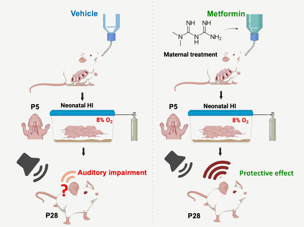
Graphic abstract. HI, hypoxia-ischemia; P5, postnatal day 5; P28, postnatal day 28.
Introduction
Neonatal hypoxia-ischemia (HI), commonly presenting clinically as hypoxic-ischemic encephalopathy (HIE), remains a leading cause of death and long-term neurodevelopmental disability in preterm and near-term infants [1]. Experimental HI models based on the Rice-Vannucci paradigm have been instrumental in defining how acute oxygen-glucose deprivation triggers evolving injury cascades that disrupt neuronal and glial maturation [2]. Because early postnatal life is a critical period for sensory circuit assembly, HI during this window can impair not only cortical and hippocampal development but also subcortical sensory pathways. In particular, emerging clinical and preclinical evidence indicates that the neonatal auditory brainstem is vulnerable to HI, positioning the auditory system as a sensitive readout of HI-related disruption in early sensory brain development [3,4].
The auditory brainstem is well suited for developmental injury studies because it comprises discrete nuclei and fast synaptic relays that mature rapidly after birth. Auditory brainstem responses (ABRs) provide a quantitative, noninvasive in vivo measure of lower auditory pathway function, capturing auditory sensitivity as well as neural synchrony and timing across brainstem relays [5]. Consistent with this, studies in neonates with HIE report ABR abnormalities including elevated thresholds, prolonged wave latencies, reduced wave V amplitude, and prolonged I–V intervals that scale with disease severity, supporting ABR as a sensitive indicator of brainstem dysfunction after perinatal HI [4,6].
Maternal metabolic state and prenatal drug exposures can shape fetal and neonatal brain development, potentially altering vulnerability to early-life insults such as HI [7,8]. Metformin is a biguanide widely prescribed for the therapy of type 2 diabetes mellitus and metabolic syndrome [9] and increasingly used during pregnancy for metabolic indications [10], and importantly it readily crosses the placenta, resulting in meaningful fetal exposure [11]. However, these safety concerns have intensified interest in its downstream effects on offspring development. Consistent with this concern, experimental and translational studies indicate that metformin can modify fetal/offspring signaling and metabolism including pathways linked to AMP-activated kinase (AMPK) and cellular energetics [12,13] although long-term neurodevelopmental consequences remain incompletely defined [14,15] and may depend on maternal context and developmental timing [16]. In parallel, metformin has been reported to improve outcomes when administered after neonatal HI in preclinical models, supporting the broader concept that metabolic modulation can influence injury evolution and repair processes in the immature brain [17,18]. However, whether maternal metformin exposure, delivered indirectly to the offspring during gestation, alters the susceptibility of developing sensory circuits to neonatal HI remains largely unknown. To address this gap, we administered metformin to pregnant dams and induced HI in pups, then assessed offspring outcomes using ABR, immunostaining of auditory brainstem, and Western blot of brain tissue to confirm metformin-associated neuroprotective effects in the offspring.
Methods
1. Animals
All animal procedures were performed in accordance with the University of Health Science Center, San Antonio Institutional Animal Care and Use Committee guidelines and complied with the National Institutes of Health Guide for the Care and Use of Laboratory Animals. Timed-pregnant Sprague-Dawley rats (The Jackson laboratory) were housed under standard conditions (12-hour light/dark cycle; ad libitum access to food and water) and allowed to deliver naturally. The day of birth was designated P0. Litters were maintained with the dam until weaning, and pups of both sexes were used unless otherwise noted. To minimize litter effects, pups were distributed across experimental groups within litters when possible, and analyses were performed with litter considered as the experimental unit where appropriate.
2. Metformin administration
Metformin was administered to pregnant dams via the drinking water. Metformin hydrochloride was dissolved in autoclaved drinking water at 1 mg/mL (0.1% w/v) in light-protected bottles. In a pregnant rat (~250–350 g) drinking ~30–50 mL/day, corresponds roughly to ~100–170 mg/kg/day [19]. Treatment began at gestational day 16 and continued until birth. Gestational day 16 was selected because it corresponds to the late-gestational period of rapid fetal brain development in the rat, allowing adequate transplacental metformin exposure prior to the neonatal hypoxic-ischemic insult. Control dams received standard drinking water prepared and handled in parallel.
3. Neonatal HI model
Postnatal HI was induced in rat pups using a unilateral carotid ligation plus systemic hypoxia paradigm adapted from the classic Rice-Vannucci model and subsequent refinements [2,20]. On postnatal day 5 (P5), pups were anesthetized with isoflurane and maintained at ~37°C. A midline neck incision was made, and left common carotid artery was carefully isolated from the vagus nerve and surrounding tissues, then permanently ligated with suture. The incision was closed using tissue adhesive, and pups were allowed to recover in a warmed environment before being returned to the dam. Sham-operated controls underwent identical anesthesia and neck exposure without carotid ligation. After recovery, pups assigned to HI were placed in a temperature-controlled, humidified hypoxia chamber maintained at ~37°C and exposed to a gas mixture containing ~8% O2 balanced with N2 at a constant flow rate for 2 hours to produce hypoxic stress in the setting of unilateral ischemia. Oxygen concentration was continuously monitored using oxygen analyzer to ensure stable hypoxic conditions. At the end of hypoxia, pups were returned to room air and placed on a warming surface until fully recovered, then returned to the dam. Pups were monitored postprocedure for general health, weight gain, and neurological signs, and any animals meeting predefined humane endpoints were excluded according to the approved protocol.
4. Auditory brainstem response
At P22–23, a time point at which the rat auditory system reaches adult-level ABR thresholds, allowing reliable and interpretable measurements of auditory function, rats were anesthetized with 4% isoflurane for induction and maintained at 2% isoflurane in oxygen (1 L/min). Adequate anesthesia was confirmed by absence of toe-pinch withdrawal reflex, and body temperature was maintained at 36°C–37°C using a feedback-controlled heating pad throughout recordings. ABR recordings were performed in a sound attenuation chamber (Med Associates, USA). Subdermal needle electrodes (Rochester Electro-Medical, USA) were placed on the top of the head, ipsilateral mastoid, and contralateral mastoid as the active, reference, and ground electrode, respectively. The signal differences in the ABRs between the vertex and the mastoid electrodes were amplified and filtered (100–5,000 Hz). Acoustic stimuli were generated by an Auditory Evoked Potentials Workstation (Tucker-Davis Technologies [TDT], USA). Closed-field click stimuli were presented monaurally to each ear. Broadband clicks (0.1- msec duration) were delivered via a 10-cm plastic tube (Tygon; 3.2-mm outer diameter) connected to TDT Multi- Field Magnetic Speakers. A total of 512 artifact-free sweeps were averaged per intensity level at a repetition rate of 16 stimuli per second. Stimulus intensities were presented in descending order from 90- to 20-dB sound pressure level (SPL) in 5-dB decrements. The left and right ears were each tested in randomized order, with additional measurements obtained near putative threshold levels; control (n=7), HI (n=6), HI-Met (n=8). Frequency-specific tone-burst stimuli (3-msec duration, 0.2-msec rise/fall times, Blackman window, alternating polarity) were generated at 8, 12, 16, and 32 kHz. Stimuli were presented in free-field configuration with the speaker positioned 10 cm from the test ear. Intensity levels ranged from 90- to 20-dB SPL in 10-dB decrements at 16 stimuli per second, with 512 artifact-free sweeps averaged per condition; control (n=4), HI (n=7), HI-Met (n=8). ABR threshold was defined as the lowest stimulus intensity producing a reproducible waveform in 2 independent averages with appropriate latency-intensity progression for identifiable waves II–V. Threshold determination was conducted by 2 blinded investigators, with third-party arbitration for disagreements. Wave amplitude measurements were obtained at standardized 85-dB SPL click stimulation; control (n=7), HI (n=7), HI-Met (n=8). Conduction time analysis included peripheral conduction time (wave I peak latency); control (n=2), HI (n=4), HI-Met (n=7) and central conduction time (wave I–IV interpeak latency); control (n=4), HI (n=4), HI-Met (n=7).
5. Immunohistochemistry
For immunohistochemistry, pups were sacrificed at group- and antigen-specific time points by deep anesthesia. At P14 and P27, the control and HI groups were examined (P14: control [n=3], HI [n=5]; P27: control [n=4], HI [n=5]). At P22–23, 3 groups (control, HI, HI-Met) were examined, with animal numbers differing by antigen (MAP2 or NeuN: control [n=12], HI [n=12], HI-Met [n=10]; OLIG1: control [n=14], HI [n=12], HI-Met [n=10]). Following decapitation, the brainstem was rapidly dissected and placed in ice-cold, low-calcium artificial cerebrospinal fluid equilibrated with carbogen (95% O2/5% CO2; 310–320 mOsm; pH 7.3–7.4). Transverse sections (100 μm) were cut on a vibratome (Leica VT1000S) and fixed in 4% paraformaldehyde in phosphate-buffered saline (PBS) for 30 minutes at 4°C. Free-floating sections were blocked in PBS containing 4% normal goat serum and 0.3% Triton X-100 for 1 hour at room temperature, then incubated overnight at 4°C with primary antibodies against MAP2 (neurons; 1:100; Sigma-Aldrich, M3696, USA or Millipore, MAB3418, Germany), NeuN (neuronal nuclei; 1:200; Millipore, MAB377), and/or OLIG1 (oligodendrocyte lineage; 1:500; Millipore, MAB5540) on separate sections as indicated. After PBS washes, sections were incubated with Alexa Fluor-conjugated secondary antibodies (1:500; Invitrogen, USA) for 2 hours at room temperature in the dark, counterstained with 4'-6-diamidino-2-phenylindole (1 μg/mL, 10 minutes), and mounted with an antifade medium (Vectashield or Fluoroprep). Images were acquired using a Zeiss LSM confocal microscope with appropriate laser lines. For each animal, analyses were performed on randomly selected fields from one section. Z-stacks were collected with specimen-dependent z-steps. For quantification, a fixed number of consecutive optical sections (~20) was analyzed per stack. Sampling volumes were calculated from voxel dimensions and field size metadata (Bio-Formats) in FIJI/ImageJ. Immunopositive cells were counted manually within each imaged field. Since the imaged area varied across sections, raw cell counts were normalized to a standardized area of 200×200 μm (40,000 μm²). Cell density was expressed as the number of immunopositive cells per 40,000 μm².
6. Western blot analysis
For Western blot analysis, pups were sacrificed on P30 by deep anesthesia (n=1 per group). Brain tissue samples were homogenized in radio immunoprecipitation assay lysis buffer supplemented with protease and phosphatase inhibitors (Halt Protease and Phosphatase Inhibitor Cocktail, Thermo Fisher Scientific, USA) and centrifuged at 13,000 rpm for 10 minutes at 4°C. Protein concentrations were measured using the BCA (bicinchoninic acid) protein assay kit (Pierce, Thermo Fisher Scientific), and equal amounts of protein were separated by sodium dodecyl sulfate-polyacrylamide gel electrophoresis on 10%–12% polyacrylamide gels. Proteins were then transferred to nitrocellulose membranes (Bio-Rad, USA). After blocking with 5% skim milk in Tris-buffered saline containing 0.1% Tween-20 for 1 hour at room temperature, membranes were incubated with primary antibodies at 4°C overnight. Primary antibodies used were: phospho-AMPKα (Thr172) (1:1000; Cell Signaling Technology, #2535, USA), AMPKα (1:1000; Cell Signaling Technology, #2532), and β-actin (1:3000; Cell Signaling Technology, #4970). After washing, membranes were incubated with horseradish peroxidase-conjugated secondary antibody (1:5000; Jackson ImmunoResearch Labs, #111-035-003, USA) for 1 hour at room temperature. Protein bands were visualized using enhanced chemiluminescence substrate (SuperSignal West Pico PLUS, Thermo Fisher Scientific) and analyzed using ImageJ software. Target protein expression levels were normalized to β-actin. For AMPK signaling analysis, both phosphorylated AMPKα and total AMPKα levels were first normalized to β-actin, and AMPK activation was subsequently expressed as the ratio of phosphorylated AMPKα to total AMPKα (p-AMPKα/AMPKα).
7. Statistical analysis
For ABR analysis, Click-evoked ABR thresholds and wave amplitudes were analyzed using 1-way analysis of variance (ANOVA) with Tukey multiple comparisons for post hoc testing, maintaining familywise error rate at α=0.05. ABR conduction time analyses employed robust statistical approaches when heteroscedasticity was detected. Both Welch ANOVA and Brown-Forsythe ANOVA were performed, with Welch ANOVA selected as the primary method due to robust performance under unequal variance conditions while maintaining appropriate type I error control. Tone-burst ABR thresholds were analyzed using linear mixed-effects models to account for repeated measures across frequencies within individual subjects. The model included group (control, HI, HI-Met) and frequency (8, 12, 16, 32 kHz) as fixed effects, with group×frequency interaction terms. Individual subjects were included as random intercepts to account for baseline hearing sensitivity differences. The model was fitted using restricted maximum likelihood estimation. Within-frequency simple effects were evaluated using Tukey adjustment for multiple comparisons, with separate familywise error correction applied for each frequency family at α=0.05. The animal was considered the primary experimental unit. All statistical analyses were conducted using GraphPad Prism ver. 11 (GraphPad Software Inc., USA). Data are presented as mean±standard error of the mean (SEM). Two-sided significance testing was employed throughout, with P<0.05 considered statistically significant.
For immunohistochemistry, differences in MAP2-positive (MAP2+) and OLIG1-positive (OLIG1+) immunopositive cell density were compared between the control and HI groups at each time point (P14 and P27) using Welch t test (P14: control [n=3], HI [n=5]. P27: control [n=4], HI [n=5]). Data are presented as mean±SEM. A P value<0.05 was considered statistically significant. AP2+/NeuN+ and OLIG1+ cell density among 3 groups (control, HI, and HIMet) at P22–23 were analyzed using the Kruskal-Wallis test, as not all groups met the assumption of normality, followed by Dunn multiple comparisons test (MAP2 or NeuN: control [n=12], HI [n=12], HI-Met [n=10]; OLIG1: control [n=14], HI [n=12], HI-Met [n=10]). Data are presented as mean±SEM, and a P value <0.05 was considered statistically significant.
Results
1. Maternal metformin treatment modulates AMPK signaling in the offspring brain following neonatal HI injury
To explore whether maternal metformin exposure during pregnancy might alter metabolic signaling in the offspring brain after neonatal hypoxic-ischemic, we examined AMPK activation in pup brain tissue at P30 by Western blot analysis (Fig. 1). Because only a single sample per group was available, these data are descriptive and were not subjected to statistical comparison. In this observation, the p‑AMPK/AMPK ratio appeared lower in the HI pup relative to control, and the sample from the metformin-treated dam (HI-Met) appeared intermediate between the control and untreated HI pup. These observations are consistent with the possibility that HI may impair AMPK activation and that maternal metformin treatment could influence this signaling. However, given the limited sample size (n=1 per group), no firm conclusions can be drawn. Rather, these preliminary findings suggest a potential link between maternal metformin exposure during pregnancy and altered metabolic signaling after neonatal HI. This observation will require confirmation in adequately powered studies.
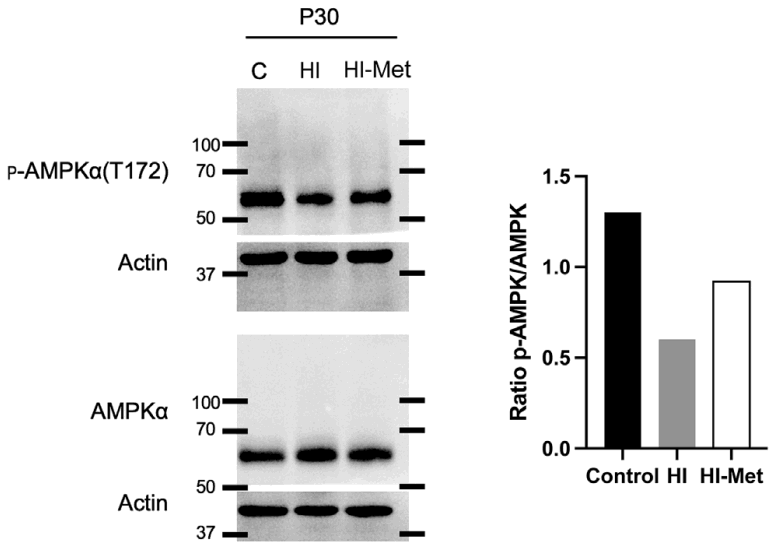
AMPK activation in offspring brain after neonatal hypoxiaischemia (HI). (A) Western blots of phosphorylated AMPKα (p-AMPKα, Thr172) and total AMPKα in brain lysates collected at postnatal day 30 (P30) from Control, HI, and HI after maternal metformin (HIMet) groups. β-Actin is shown as a loading control for each blot. (B) p-AMPKα/total AMPKα ratio. Relative to control, the p-AMPKα/AMPKα ratio appeared lower in the HI sample and intermediate in the HI-Met sample; as only a single sample per group was analyzed, these values were not statistically compared (n=1 per group). AMPK, AMP-activated kinase.
2. Maternal metformin recovers HI-associated elevations in ABR threshold and preserves brainstem response amplitudes
To determine whether maternal metformin exposure during pregnancy modifies offspring auditory dysfunction after neonatal HI, we recorded in vivo ABRs in control, HI, and HI-Met groups at P22–23. Representative traces showed delayed peak timing and reduced response amplitudes in the HI group relative to controls, whereas waveforms from HI-Met pups more closely resembled control responses (Fig. 2A).
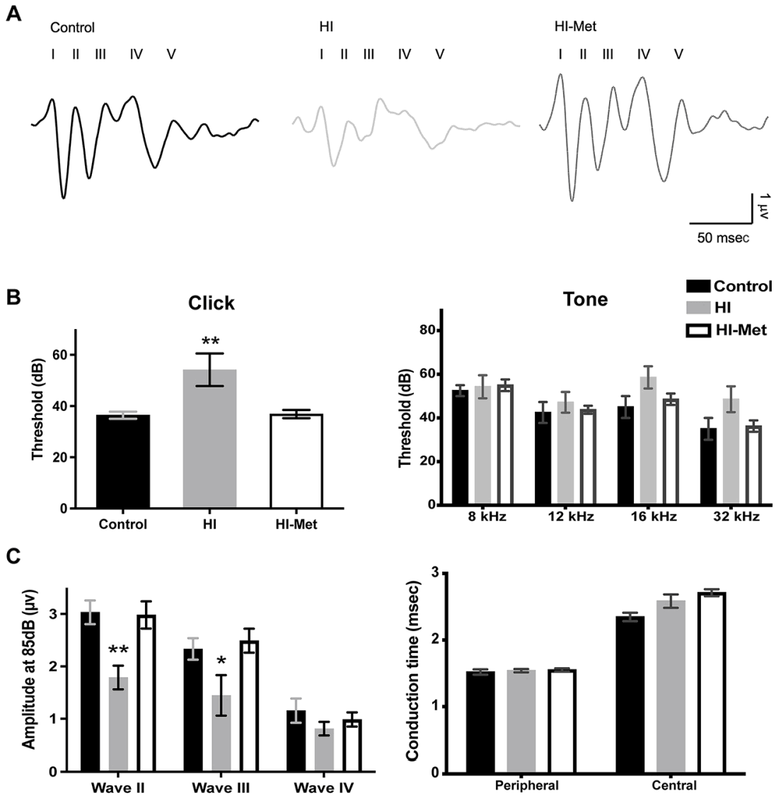
Therapeutic effects of maternal metformin on auditory brainstem responses following neonatal hypoxia-ischemia (HI). (A) Representative click-evoked auditory brainstem response (ABR) waveforms recorded at 85-dB sound pressure level (SPL) from control (black), HI (gray), and HI after maternal metformin (HI-Met; white) groups, with peaks I–V annotated. Scale bars: 1 μV (vertical), 50 msec (horizontal). The HI group shows reduced wave II and III amplitudes relative to control, while the HI-Met group preserves waveform morphology comparable to control. (B) Group summary data for click-evoked ABR thresholds (left panel) and tone-burst ABR thresholds at 8, 12, 16, and 32 kHz (right panel). Click-evoked thresholds differed significantly among groups (1-way ANOVA: F [2,18]=8.132, df=0.003; control [n=7], HI [n=6], HI-Met [n=8]). Tone-burst thresholds showed no significant group differences at any tested frequency (mixed-effects model with Tukey-adjusted comparisons; all Tukey-adjusted P>0.05; control [n=4], HI [n=7], HI-Met [n=8]). (C) Wave II–IV amplitudes at 85-dB SPL (left panel) and click-evoked ABR conduction times (right panel), including peripheral (wave I latency) and central (I–IV interpeak latency) measures. No significant group differences were observed in wave amplitudes or conduction times, indicating preserved neural transmission timing across groups (mixed-effects model with Tukey correction; control [n=7], HI [n=7], HI-Met [n=8]; variance-robust ANOVA for conduction times; Peripheral conduction times: control [n=2], HI [n=4], HI-Met [n=7], total [n=13], central conduction times: control [n=4], HI [n=4], HI-Met [n=7], total [n=15]). Data are presented as mean±standard error of the mean. *P<0.05, **P<0.01 after post hoc adjustment. ANOVA, analysis of variance.
Consistent with these qualitative differences, HI significantly impaired auditory sensitivity, producing an elevation in click-evoked ABR threshold that was prevented by maternal metformin exposure (control: 36.43±1.43-dB SPL; HI: 54.17±6.38-dB SPL; HI-Met: 36.88±1.62-dB SPL; 1-way ANOVA: F [2,18]=8.132, P=0.003; control [n=7], HI [n=6], HI-Met [n=8]; Fig. 2B, left panel). Post hoc analysis using Tukey multiple comparisons revealed that HI groups had significantly elevated thresholds compared to both control and HI-Met groups (HI exceeded control by 17.74 dB; 95% confidence interval [CI] for control-HI: -30.50 to -4.979; Tukey-adjusted P=0.006; HI exceeded HI-Met by 17.29 dB; 95% CI, 4.906–29.68; Tukey-adjusted P=0.006). Importantly, no significant difference was observed between control and HI-Met groups (95% CI, -12.32 to 11.42; Tukey-adjusted P=0.995), indicating complete prevention of HI-induced hearing threshold elevation with metformin treatment.
Tone-burst thresholds (8–32 kHz) showed the same directionality but did not reach statistical significance after correction for multiple comparisons. Using a mixed-effects model assessing group×frequency with within-frequency simple effects (Tukey-adjusted alpha=0.05; control [n=4], HI [n=7], HI-Met [n=8]), no group differences were detected at 8 or 12 kHz. At 16 and 32 kHz, HI pups showed nonsignificant trends toward higher thresholds compared with controls (16 kHz: mean difference for control-HI=-13.57 dB; 95% CI, -33.90 to 6.758; Tukey-adjusted P=0.199; 32 kHz: mean difference for control-HI=-13.57 dB; 95% CI, -35.38 to 8.240; Tukey-adjusted P=0.243), while HI-Met thresholds remained comparable to controls across frequencies (Fig. 2B, right panel). Together, these data demonstrate a robust HI-induced elevation in click threshold at P22–23 and suggest a tendency toward high-frequency vulnerability, while maternal metformin exposure preserves auditory sensitivity after HI, consistent with functional protection of the developing auditory pathway.
We next quantified ABR wave amplitudes as a functional readout of neural synchrony and relay strength across the auditory brainstem. At a standardized 85-dB click intensity, wave II and wave III amplitudes were significantly reduced in the HI group compared with both control and HI-Met groups (wave II: P<0.01; wave III: P<0.05, Tukey-adjusted; control n=7, HI n=7, HI-Met n=8; Fig. 2C, left panel). Notably, wave II and wave III amplitudes did not differ between control and HI-Met pups, indicating that maternal metformin exposure preserved these brainstem response components. Wave IV amplitude did not differ significantly among groups under Tukey-adjusted testing, suggesting that HI selectively disrupted specific ABR components rather than producing a uniform reduction across all waves.
To determine whether HI alters transmission speed along the auditory pathway, we also analyzed conduction metrics at 85 dB. Peripheral conduction time, defined as wave I peak latency, was indistinguishable among control, HI, and HI-Met groups (Welch ANOVA; W=0.1665, df=2, 3.087, P=0.854 and Brown-Forsythe ANOVA; F*=0.217, df=2, 4.753, P=0.813; control [n=2], HI [n=4], HI-Met [n=7], total [n=13]). Central conduction time, measured as the wave I to wave IV interpeak latency at 85 dB, also demonstrated no significant among-group differences (Welch ANOVA; W=1.950, df=2, 5.443, P=0.230 and Brown-Forsythe ANOVA; F*=2.046, df=2, 7.465, P=0.196; control [n=4], HI [n=4], HIMet [n=7], total [n=15]; Fig. 3C, right panel). Together, these data indicate that neonatal HI primarily affected hearing sensitivity and reduced neural response magnitude without detectably slowing auditory nerve or brainstem conduction, and that maternal metformin exposure preserved suprathreshold ABR amplitudes while maintaining normal conduction properties.
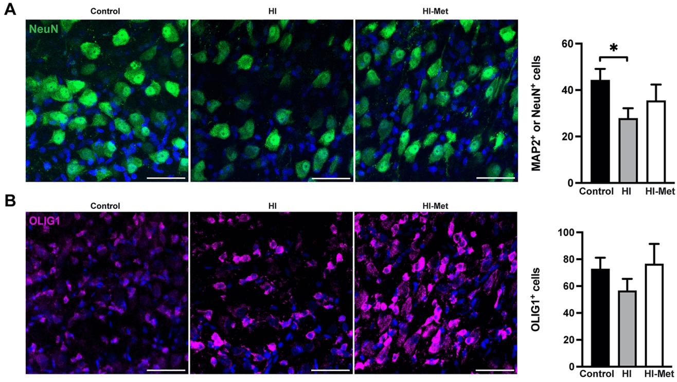
Effects of maternal metformin on neuronal and oligodendrocyte lineage cell density at P27 following neonatal hypoxia-ischemia (HI). (A) Representative images and quantification of MAP2-positive (MAP2+) or NeuN-positive (NeuN+) neuronal density (the number of cells per 40,000 μm2) in brainstem sections from control (black), HI (gray), and HI after maternal metformin (HI-Met; white) groups. The HI group showed significantly reduced neuronal density compared to control (*P<0.05; control [n=12], HI [n=12], HI-Met [n=10]), while the HI-Met group showed an intermediate density that did not differ significantly from either group. (B) Representative images and quantification of OLIG1-positive (OLIG1+) oligodendrocyte lineage cell density. No significant differences were observed among the 3 groups (control [n=14], HI [n=12], and HI-Met [n=10]). Data are presented as mean±standard error of the mean. Statistical comparisons among groups were performed using the Kruskal-Wallis test with Dunn post hoc multiple comparisons. Scale bars: 50 μm. *P<0.05.
3. HI induces delayed neuronal loss and dynamic oligodendrocyte lineage responses in the MNTB
Because ABR wave II and wave III amplitudes, indices of neural synchrony and relay strength in the auditory brainstem, were significantly reduced in HI pups, we next asked whether HI produces corresponding cellular pathology in auditory brainstem nuclei. We focused on the medial nucleus of the trapezoid body (MNTB), a key relay within the sound-localization pathway, and quantified neuronal and oligodendrocyte lineage populations by immunohistochemistry at 2 postinjury time points. To define the developmental trajectory of HI-associated changes, we first assessed MAP2+ neurons and OLIG1+ oligodendrocyte lineage cells at postnatal day P14 and P27.
MAP2+ neuronal density exhibited an age-dependent response to HI. At P14, MAP2+ cell counts were numerically higher in HI pups: 28.22±4.41 cells/40,000 μm² than in controls:19.22±0.33 cells/40,000 μm², although this difference did not reach statistical significance (Welch t test, t=2.031, P=0.111; control [n=3], HI [n=5]). By P27, this pattern reversed, with a pronounced and significant reduction in MAP2+ neurons in the HI group: 3.34±0.52 cells/40,000 μm² compared with controls: 15.42±1.61 cells/40,000 μm² corresponding to a ~78% decrease in neuronal density (Welch t test, t=7.158, P=0.0029; control [n=4], HI [n=5], Fig. 4A). These findings indicate that HI is associated with minimal or variable changes in MAP2+ labeling early after injury, but substantial neuronal loss emerges at later postnatal stages.
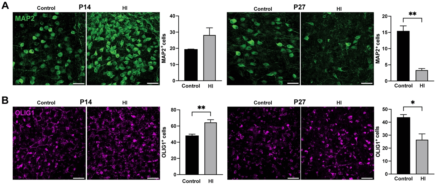
Temporal changes in neuronal and oligodendrocyte lineage cell density following neonatal hypoxia-ischemia (HI). (A) Representative images and quantification of MAP2-positive (MAP2+) neuronal density (the number of cells per 40,000 μm2) in brainstem sections from control and HI animals at P14 and P27. MAP2+ cell density was significantly reduced in HI animals at P27 (**P<0.01; control [n=4], HI [n=5]), but not at P14 (df=0.111; control [n=3], HI [n=5]). (B) Representative images and quantification of OLIG1-positive (OLIG1+) oligodendrocyte lineage cell density at P14 and P27. OLIG1+ density was significantly elevated in HI animals at P14 (**P<0.01; control [n=3], HI [n=5]) and significantly reduced at P27 (*P<0.05; control [n=4], HI [n=5]). Data are presented as mean±standard error of the mean. Comparisons between control and HI groups at each time point were performed using Welch unpaired t test. Scale bars: 50 μm. *P<0.05, **P<0.01. P14, postnatal day 14; P27, postnatal day 27.
A parallel but contrasting temporal pattern was observed in OLIG1+ oligodendrocyte lineage cells. At P14, OLIG1+ cell density was significantly elevated in HI pups (64.41±3.46 cells/40,000 μm²) compared with controls (47.79±2.17 cells/40,000 μm²; Welch t test, t=4.072, P=0.007; control [n=3], HI [n=5], Fig. 4B), suggesting an early reactive expansion of the oligodendrocyte lineage in response to hypoxic-ischemic injury. By P27, however, this initial increase was reversed, with OLIG1+ density falling significantly below control levels in the HI group (26.49±4.55 cells/40,000 μm² vs. 43.54±2.35 cells/40,000 μm²; Welch t test, t=3.328, P=0.016; control [n=4], HI [n=5]), indicating progressive depletion of the oligodendrocyte lineage pool over time. Taken together, the bidirectional temporal responses of both MAP2+ neurons and OLIG1+ oligodendrocyte lineage cells suggest that neonatal HI initiates a dynamic, time-dependent process of cellular injury in the brainstem, with early reactive changes giving way to substantial cell loss by the third postnatal week.
Thus, these results suggest progressive vulnerability of MNTB cellular populations following HI, with delayed and robust neuronal loss and a more variable oligodendrocyte lineage response. This alteration provides a plausible cellular substrate for the reduced ABR wave amplitudes observed after HI.
4. Maternal metformin preserves MNTB neuronal populations after neonatal HI
To test whether maternal metformin exposure during pregnancy modifies HI-associated cellular pathology in the developing auditory brainstem, we quantified neuronal and oligodendrocyte lineage populations in the MNTB at P22–23 across control, HI, and HI-Met groups. Neuronal density was assessed using alternating sections stained for MAP2 (morphologically intact neurons) or NeuN (nuclear-intact neurons) and counts from the 2 markers were averaged for each animal. This analysis revealed a significant overall group effect (Kruskal-Wallis test, H [2]=7.827, P=0.020; control [n=12], HI [n=12], and HIMet [n=10]). Post hoc Dunn multiple comparisons test demonstrated that neuronal density was significantly lower in the HI group compared with controls (adjusted P=0.018), while the HI-Met group did not differ significantly from control (adjusted P=0.228). No significant difference was observed between the HI and HI-Met groups (adjusted P>0.999), suggesting partial preservation of neuronal density with metformin treatment. Compared with controls (44.30±4.83 cells/40,000 μm²), HI produced a marked reduction in neuronal density (27.81±4.37 cells/40,000 μm²; ~37% decrease). Importantly, HI-Met pups exhibited partial preservation of neuronal populations after HI (35.43±6.92 cells/40,000 μm², Fig. 3A), corresponding to ~46% recovery toward control levels, consistent with a neuroprotective effect of maternal metformin exposure during this postinjury developmental window.
In parallel, OLIG1+ oligodendrocyte lineage cell density showed a similar directional pattern, although the overall group effect did not reach statistical significance (Kruskal-Wallis test, H [2]=2.034, P=0.362; control [n=14], HI [n=12], HI-Met [n=10]). Mean OLIG1+ counts were 72.7±8.40 cells/40,000 μm² in controls, 56.48±8.92 cells/40,000 μm² in HI pups, and 76.47±14.90 cells/40,000 μm² in HI-Met pups (Fig. 3B). Post hoc Dunn multiple comparisons indicated a numerical reduction in OLIG1+ cells after HI relative to controls (adjusted P=0.497) and a trend toward preservation with maternal metformin exposure (HI vs. HI-Met: adjusted P=0.985). Together, these findings suggest that maternal metformin administration mitigates HI-induced loss of neuronal markers in the MNTB and may help maintain oligodendrocyte lineage populations, supporting a broader protective influence on cellular substrates relevant to auditory brainstem function.
Discussion
This study demonstrates that maternal metformin during pregnancy attenuates neonatal HI-induced hearing loss in offspring. Rat pups from the metformin-treated group showed ABR thresholds and wave amplitudes comparable to controls. Notably, auditory function showed substantial recovery even though cellular protection was incomplete, suggesting that maternal metformin exposure may support functional preservation of the auditory pathway through mechanisms that extend beyond structural rescue.
1. Gestational metformin preserves auditory brainstem function despite partial cellular recovery and age-dependent vulnerability
Offspring from metformin-treated dams exhibited robust functional protection after neonatal HI, including complete normalization of click-evoked ABR thresholds, full preservation of wave II and III amplitudes, reflecting the cochlear nucleus and the superior olivary complex (SOC), and normal peripheral and central conduction times. This functional preservation was accompanied by partial rescue of MNTB neuronal integrity within the SOC circuitry, with MAP2+ neuronal counts recovering substantially toward control levels in the HI-Met group compared with HI. Notably, the observation that ABR function was fully normalized despite only partial restoration of neuronal and glial populations is consistent with the concept of neural redundancy, in which surviving elements and preserved connectivity can be sufficient to maintain circuit-level output [21,22]. In this context, metformin may preferentially protect functionally critical neuronal subpopulations, stabilize synaptic transmission, and/or preserve myelin-dependent conduction fidelity, thereby sustaining auditory brainstem performance.
In parallel, the divergent cellular profiles across development highlight an age-dependent trajectory of vulnerability in the auditory brainstem. While MAP2+ neuronal labeling showed little change at P14, a pronounced loss emerged by P27, indicating delayed neuronal vulnerability during later postnatal maturation. OLIG1+ oligodendrocyte lineage cells followed a similarly dynamic pattern, with a trend toward increased density at P14, potentially reflecting reactive expansion or altered differentiation state, followed by reduced density at later time points (P27 and P22–23), consistent with progressive depletion and/or impaired maturation of the oligodendrocyte lineage after HI. Importantly, maternal metformin exposure appeared to maintain OLIG1+ cell numbers near control levels at P22–23, supporting the idea that gestational metformin confers cellular resilience in the developing brainstem, potentially through preservation of oligodendroglial populations and white matter integrity during a critical window of postnatal circuit refinement.
2. Gestational metformin and metabolic signaling after neonatal HI
Our exploratory Western blot analysis, the phosphorylation ratio (p-AMPKα [Thr172]/total AMPKα) at P30 appeared lower in the HI offspring than in the control, and the sample from a metformin-treated dam appeared intermediate. These data are insufficient to conclude that neonatal HI suppresses, or that gestational metformin preserves, AMPK activation in the developing brain.
AMPK phosphorylation coordinates cellular energy stress responses, initiating autophagy, mitochondrial quality control, and bioenergetic adaptation [23]. In neurons, AMPK phosphorylation facilitates mitophagy activation and metabolic adaptation during energy stress [24]. The spiral ganglion neurons and brainstem auditory nuclei are highly energy demanding brain regions exhibiting a high NAD(P)H/FAD turnover during sustained or high-frequency firing [25,26]. Because AMPK is a central regulator of cellular energy homeostasis, maintenance of AMPK signaling may enhance metabolic adaptability in injured neonatal brain tissue and support recovery processes during ongoing postnatal maturation. However, this study has limitations. We did not directly measure metformin concentrations in fetal or neonatal brain tissue, and AMPK outcomes were assessed at a single time point in a single sample per group. Adequately powered, longitudinal profiling of metformin exposure and AMPK signaling across developmental stages will be necessary before any conclusions about the durability or timing of such effects can be drawn.
Maternal metformin exposure normalized click-evoked ABR thresholds and preserved suprathreshold wave amplitudes without altering conduction times, consistent with maintained circuit output rather than generalized changes in transmission speed. In parallel, metformin partially rescued HI-associated reductions in neuronal markers within the MNTB and showed a trend toward preserving OLIG1+ oligodendrocyte lineage cells. While these functional and cellular results are consistent with a protective effect on vulnerable auditory brainstem circuits following neonatal HI, the proposed link to AMPK-mediated metabolic resilience remains a hypothesis that requires further mechanistic studies.
3. Clinical implications
Therapeutic hypothermia remains the only established therapy for moderate-to-severe neonatal HIE, yet composite adverse outcomes (death or severe disability) occur in 38-59% of cases [27,28], underscoring the urgent need for complementary neuroprotective strategies. In this context, maternal metformin represents a preventive strategy complementary to postnatal therapies. A recent study has proposed postnatal metformin administration (32 mg/kg/day) in HIE infants based on physiologically based pharmacokinetic modeling [29]. Preclinical studies demonstrate neurorestorative effects including neural precursor cell activation [30], improved remyelination and oligodendrocyte progenitor cell proliferation [31], and neuroprotection through AMPK activation and anti-inflammatory mechanisms [32]. Maternal administration offers earlier intervention before injury and avoids direct drug administration to vulnerable neonates. These dual strategies—maternal prevention and postnatal regeneration—may optimize neuroprotection across HIE injury phases. In addition, the low cost and accessibility of metformin may be particularly relevant for low- and middle-income countries, where HIE incidence remains high and access to therapeutic hypothermia infrastructure is limited [33]. In resource-limited settings with a high burden of birth asphyxia-related neonatal morbidity and limited access to therapeutic hypothermia, maternal metformin could provide a low-cost, widely accessible preventive intervention that does not require specialized neonatal intensive care infrastructure.
Nevertheless, these potential benefits must be weighed against safety considerations. In the present study, maternal and offspring safety parameters were not systematically recorded. Because metformin crosses the placenta and in utero exposure has been associated with changes in offspring development and metabolic function, additional safety studies evaluating the effects of gestational metformin exposure are warranted before clinical translation [34].
In conclusion, maternal metformin administration during pregnancy was associated with functional protection of the developing auditory pathway following neonatal HI, accompanied by partial cellular rescue. Together, the preserved ABR function and partial histological rescue are consistent with a model in which maternal metformin supports the maintenance of vulnerable auditory brainstem circuits after injury. Although our findings support the potential protective effect of maternal metformin exposure, future studies may need to examine whether acute maternal administration of metformin shortly before delivery is effective in neonatal HI injury animal models and including the incorporation of Sham groups to assess effects on normal pups. Ultimately, carefully designed studies would help define the therapeutic window, feasibility, and translational relevance of this approach before clinical trials.
Notes
Conflicts of interest
No potential conflict of interest relevant to this article was reported.
Funding
This study received no specific grant from any funding agency in the public, commercial, or not-for-profit sectors.
Author contribution
Conceptualization: SEK, YJA, JHK; Formal Analysis: SEK, JYK, YJA, JHK; Investigation: SEK, JYK, YJA; Methodology: SEK, YJA, JHK; Project Administration: SEK, JHK; Writing – Original Draft: SEK, JYK, YJA, JHK; Writing – Review & Editing: SEK, JYK, YJA, JHK