Evidence-based clinical practice guidelines for pediatric hepatoblastoma: a systematic review and expert recommendations from Korea
Article information

Abstract
Hepatoblastoma (HB) is the most common primary liver malignancy in children, predominantly affecting those younger than 3 years of age. Although the overall survival rate has improved significantly over the past few decades, standardized clinical practice guidelines for HB in Korea have been lacking. This clinical practice guideline was developed to provide evidence-based recommendations for the diagnosis and treatment of HB in children. A multidisciplinary working group comprising pediatric hematologists/oncologists, radiologists, surgeons, pathologists, and methodologists systematically reviewed the literature published up to May 2023. For pretreatment imaging, both abdominal computed tomography and magnetic resonance imaging are useful for PRE-Treatment EXTent of disease (PRETEXT) staging, with each modality offering distinct advantages. PRETEXT staging at diagnosis is recommended as it provides valuable prognostic information. For standard-risk HB (PRETEXT I–III without metastasis and alpha-fetoprotein ≥100 ng/mL), cisplatinbased chemotherapy without doxorubicin is recommended over cisplatin-doxorubicin combination therapy, as it demonstrates equivalent survival outcomes with fewer adverse effects. In carefully selected patients who are eligible for complete resection at diagnosis, upfront surgery without neoadjuvant chemotherapy should be considered to spare unnecessary chemotherapy and its toxicities. For patients with residual pulmonary metastases after chemotherapy, pulmonary metastasectomy should be considered. Sodium thiosulfate administration following cisplatin should be considered to reduce ototoxicity, although the supporting evidence is derived predominantly from standard-risk patients and its availability in Korea remains limited. Regarding histological subtypes, well-differentiated fetal HB is associated with favorable outcomes and may be treated with surgery alone, whereas the small cell undifferentiated pattern is no longer classified as a HB subtype and requires immunohistochemical and molecular testing to distinguish it from malignant rhabdoid tumor and other small round cell tumors. This guideline represents the first evidence-based clinical practice guideline for pediatric HB in Korea. It aims to standardize the diagnostic and therapeutic approaches, ultimately improving outcomes for Korean children with HB. Given the rarity of this tumor, international collaboration and prospective data collection remain essential for future guideline updates.
Key message
This is the first Korean evidence-based clinical practice guideline for pediatric hepatoblastoma. Key recommendations include computed tomography and magnetic resonance imaging for PRETEXT (PRE-Treatment EXTent of disease) staging, cisplatin monotherapy for standard-risk disease, upfront surgery for resectable tumors, metastasectomy for residual lung lesions, and sodium thiosulfate for otoprotection. Well-differentiated fetal histology carries a favorable prognosis, whereas the small cell undifferentiated pattern requires immunohistochemical and molecular testing to exclude malignant rhabdoid tumor.
Introduction
Hepatoblastoma (HB) is the most prevalent primary hepatic malignancy in the pediatric population, accounting for over 90% of liver cancers in children under 5 years of age. The estimated annual incidence is approximately 1.5 cases per million children, which has shown a gradual increase over the past 2 decades—largely attributed to enhanced survival rates among premature and low-birthweight neonates [1].
The disease predominantly affects children younger than 3 years and typically manifests as a palpable abdominal mass accompanied by markedly elevated serum alpha-fetoprotein (AFP) levels. Prognosis depends on several key factors: the feasibility of complete surgical resection, the presence of metastatic disease, and the tumor’s histopathological characteristics.
Current therapeutic strategies are based on risk-adapted, multimodal approaches informed by prognostic indicators. Four major international cooperative groups —Children’s Oncology Group (COG), International Childhood Liver Tumours Strategy Group (SIOPEL), German Society for Pediatric Oncology and Hematology (GPOH), and Japanese Study Group for Pediatric Liver Tumors (JPLT)—have played pivotal roles in shaping contemporary treatment paradigms. These protocols primarily incorporate cisplatin (CDDP)-based chemotherapy, which has substantially improved overall survival (OS) rates for HB overall from approximately 30% to 70%–80%. Outcomes nonetheless differ substantially by risk group, exceeding 90% in completely resected standard-risk disease while remaining considerably lower in high-risk or metastatic disease.
In particular, event-free survival (EFS) in low-risk patients can now approach or exceed 90% [2], reflecting remarkable advances in both systemic therapy and surgical techniques. Notably, the integration of liver transplantation (LT) as a curative option for unresectable or advanced tumors has further contributed to improved outcomes [2].
However, there remain notable differences among the major study groups in terms of chemotherapeutic regimens, timing of surgical intervention, and overall treatment strategies. To date, no globally standardized clinical protocol has been established for the management of HB. As a result, there has been a growing consensus on the need for localized clinical guidelines that can be adapted to the specific medical environment in Korea—guidelines that encompass not only diagnosis and treatment, but also long-term follow-up after therapy completion.
This newly developed clinical practice guideline (CPG) aims to address that need by offering evidence-based, state-of-the-art treatment recommendations that can be applied uniformly across the country. Ultimately, it seeks to ensure that all pediatric patients with HB in Korea have equitable access to optimal care, thereby improving survival outcomes while also preserving quality of life during and after treatment.
Methods
1. Guideline Development Committee
In February 2022, the National Cancer Center and the Korean Society of Pediatric Hematology and Oncology initiated a joint project to develop the first Korean evidence-based CPGs for HB. A multidisciplinary guideline development committee was established, comprising a steering committee (10 members) and a working group (12 members). The committee included experts in pediatric oncology, hepatobiliary surgery, radiology, and pathology, as well as expert methodologists. The steering committee also served as a conflict of interest committee, and all members who participated in the development of the guidelines disclosed their interests before and during the development of the recommendations, and conflict of interest management standards were established. A detailed list of the guideline development committee and declarations of conflict of interest are presented in Supplementary Table 1.
2. Purpose and scope of developing CPG
The purpose of this CPG is to provide an evidence-based framework for the diagnosis and treatment of pediatric HB, enabling clinicians to make informed decisions that improve treatment outcomes and quality of life. It addresses radiologic and pathologic diagnostic methods, chemotherapeutic regimens, and surgical treatment strategies, and is intended for physicians in primary, secondary, and tertiary medical institutions who manage pediatric HB. The recommendations apply to patients from birth to 19 years of age with a confirmed diagnosis of HB.
3. Selection of key questions
The CPG was created using standard approaches for the development of evidence-based CPGs [3], employing the AGREE II (Appraisal of Guidelines for Research and Evaluation II) instrument as a framework to ensure methodological rigor and transparency [4]. The committee members developed the key questions and identified and rated the importance of outcomes by consensus.
4. Literature search and selection
The literature search strategy was developed through discussions between methodological experts and the CPG committee members responsible for each key question. Initial search terms were generated and adapted to construct search strings for MEDLINE (PubMed). These were subsequently translated into the controlled vocabulary of each target database to develop database-specific search strategies. No restrictions were applied with respect to publication year, language, or publication status at the search stage. At the screening stage, however, eligibility was restricted to articles published in English or Korean. The retrieved records were aggregated using EndNote, and duplicates were removed by matching titles, author names, publication years, and journal names. In cases where discrepancies in indexing terms between databases resulted in unmatched duplicates, manual de-duplication was performed. The databases searched included MEDLINE, Embase, the Cochrane Library, and KoreaMed. Searches were conducted between March 24, 2023, and May 4, 2023; accordingly, the evidence base of this guideline comprises literature published up to May 2023, and no updated or supplementary search was performed thereafter. For each key clinical question, at least 2 independent committee members were assigned to review and select studies, with discrepancies resolved through discussion. The study selection process was documented following the PRISMA (Preferred Reporting Items for Systematic Reviews and Meta-analyses) flow diagram, and detailed selection procedures are provided in the Supplementary Fig. 1. Inclusion and exclusion criteria were defined for each key question using the PICOS framework (Population, Intervention, Comparator, Outcomes [benefits and harms], Study design). Considering the limited availability of high-level evidence in this field, no restrictions were applied regarding study design. Non-comparative studies were also included to maximize the breadth of relevant data available for synthesis and presentation.
5. Evidence appraisal, synthesis, and development of recommendations
The quality of evidence was evaluated using the revised Cochrane risk-of-bias tool for randomized trials (ROB 2.0) [5] for articles reporting randomized studies and the Risk of Bias Assessment Tool for Non-Randomized Studies ver. 2.0 (ROBANS 2.0) [6] for articles reporting nonrandomized studies. A meta-analysis of the selected articles was performed using RevMan (ver. 5.3.3; the Nordic Cochrane Centre, Denmark). When a direct comparison between the intervention and the comparison was not feasible, a meta-analysis was not conducted; instead, a qualitative synthesis of the evidence was performed. The strength of recommendations and level of evidence were determined using the Grading of Recommendations Assessment, Development, and Evaluation (GRADE) [7] (Supplementary Table 2). The level of evidence was rated as high, moderate, low, or very low based on certainty in treatment effects for the target population. Certainty was influenced by limitations in study design and consistency, precision, and directness of the data. Recommendations were classified as either strong or weak. A strong recommendation was made when the anticipated benefits of an intervention clearly outweighed its potential risks; therefore, most patients should (or should not) receive the intervention as standard practice. A weak recommendation was made when the balance between benefits and risks was uncertain or closely matched, necessitating individualized clinical decision-making that incorporates patient and family preferences. In developing each recommendation, the committee considered not only clinical efficacy and safety but also cost-effectiveness, resource implications, and the feasibility of implementation across diverse healthcare settings.
6. Review and approval
The recommendation grading process was approved if >80% of the working group participated and >70% endorsed the recommendation. Voting was conducted during working committee meetings using a 5-point Likert scale, and adoption required ≥70% of responses as “completely agree” or “generally agree.”
7. External review
External review by clinical experts in relevant fields was requested during a public hearing on the draft recommendations. The opinions from external experts were reviewed and incorporated as appropriate in revising the draft recommendations. The final level of agreement was subsequently confirmed through a structured survey of the advisory committee members.
8. Dissemination of clinical guidelines and update plan
To ensure broad dissemination, the Clinical Practice Guidelines for Pediatric Hepatoblastoma was published as a book in July 2025. These guidelines will also be made available on the official website of the Korean Cancer Management Guideline Network (www.ko-amguide.com), and distributed through various channels. If a revision is deemed necessary, the Korean Society of Pediatric Hematology and Oncology will revise this guideline approximately every 3 years.
Results
A total of 8 recommendations were formulated to address 7 key questions pertaining to the radiologic and pathologic diagnosis, as well as chemotherapeutic and surgical management strategies for HB. The recommendations are summarized in Table 1 and in the summary figure (Fig. 1). The systematic review included 39 publications; some publications informed more than one KQ, and the flow diagrams outlining study identification and selection for each key question are provided in Supplementary Fig. 1. The GRADE summary of findings for each key question is provided in Supplementary Table 3(1–7).
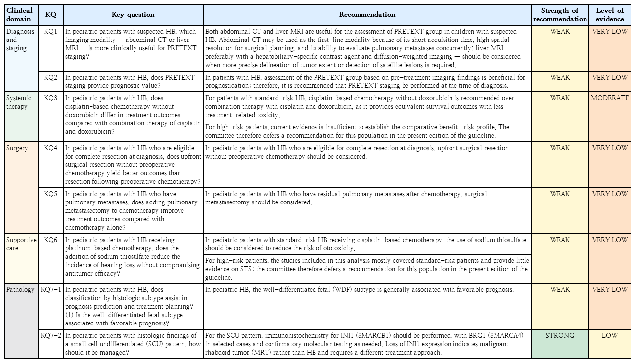
Summary of recommendations of the 2025 Korean evidence-based clinical practice guideline for the diagnosis and treatment of pediatric hepatoblastoma, developed by the Korean Society of Pediatric Hematology and Oncology and the National Cancer Center of Korea
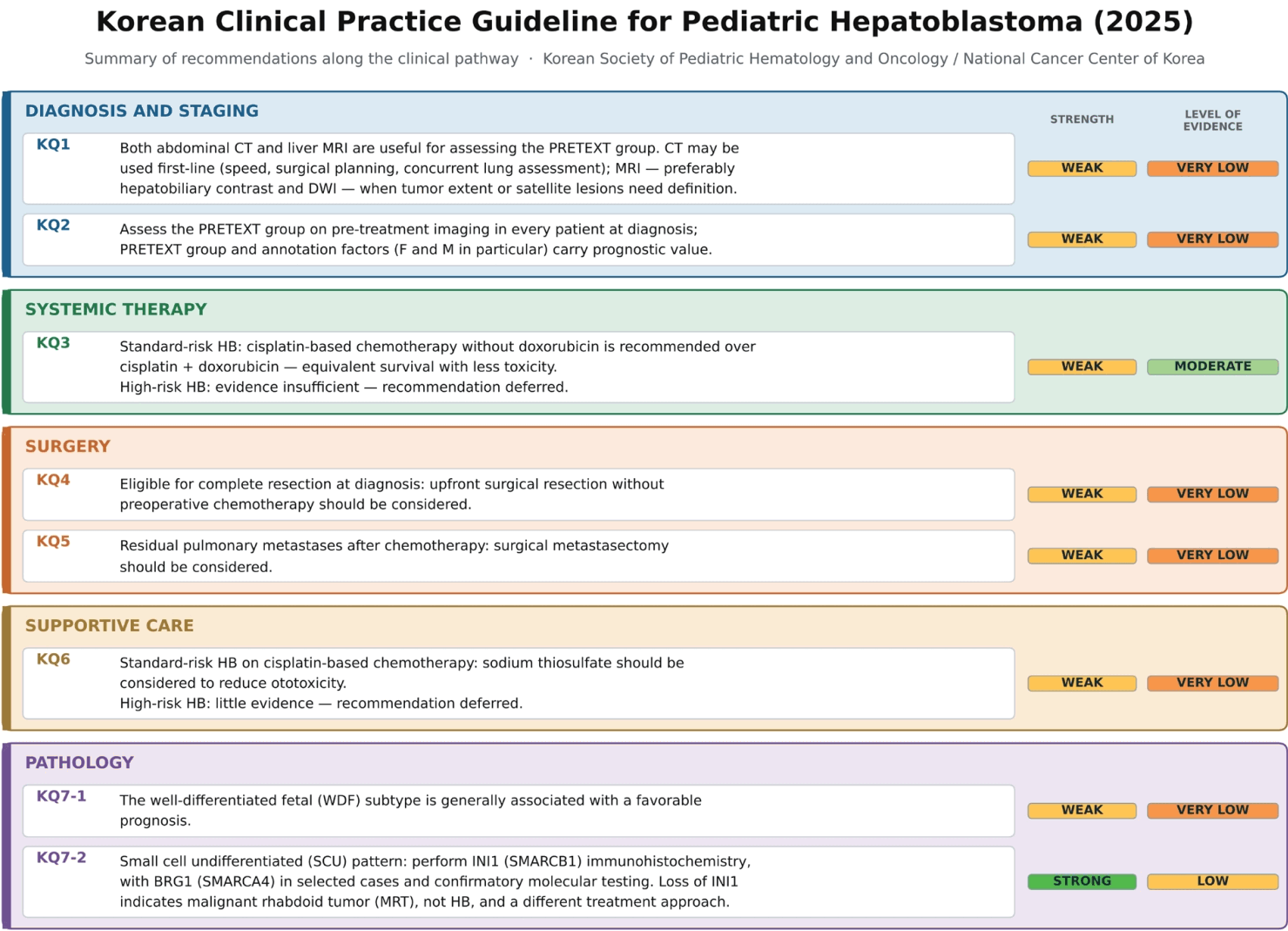
Summary of the 2025 Korean evidence-based clinical practice guideline for pediatric hepatoblastoma: recommendations along the clinical pathway, developed by the Korean Society of Pediatric Hematology and Oncology and the National Cancer Center of Korea. The 8 recommendations addressing the 7 key questions are grouped into 5 clinical domains-diagnosis and staging, systemic therapy, surgery, supportive care, and pathology. For each recommendation, the strength of recommendation (green, strong; yellow, weak) and the level of the evidence (green, high; light green, moderate; yellow, low; orange, very low) are shown according to the GRADE approach. Systematic literature searches were completed in May 2023. KQ, key question; CT, computed tomography; PRETEXT, PRE-Treatment EXTent of disease; MRI, magnetic resonance imaging; DWI, diffusion-weighted imaging; HB, hepatoblastoma; IHC, immunohistochemistry.
1. Key question 1
In pediatric patients with suspected HB, which imaging modality—abdominal computed tomography (CT) or liver magnetic resonance imaging (MRI)—is more clinically useful for PRETEXT staging?
1) Recommendation
Both abdominal CT and liver MRI are useful for the assessment of PRETEXT group in children with suspected HB. Abdominal CT may be used as the first-line modality because of its short acquisition time, high spatial resolution for surgical planning, and its ability to evaluate pulmonary metastases concurrently; liver MRI—preferably with a hepatobiliary-specific contrast agent and diffusion-weighted imaging—should be considered when more precise delineation of tumor extent or detection of satellite lesions is required. (weak recommendation, very low level of evidence)
2) Explanation
A total of 377 nonduplicate records were identified through the literature search. Titles and abstracts were screened, resulting in 27 full-text articles assessed for eligibility according to predefined inclusion and exclusion criteria. Ultimately, 2 studies were selected for analysis [8,9]. Both were pre–post studies, precluding quantitative synthesis or meta-analysis. Each was a single-center study conducted in India [9] and the United States [8], respectively. While both studies reported the impact of abdominal CT or liver MRI on tumor detection rates and PRETEXT staging, neither provided diagnostic accuracy metrics such as sensitivity or specificity.
Risk of bias was assessed using the ROBANS 2.0. The overall certainty of evidence began at the “low” level for all outcomes. In the 2022 study by Sharma et al. [9], the time interval between CT and MRI examinations was not specified, leaving open the possibility that temporal changes could have influenced results; therefore, the risk-of-bias rating was downgraded for potential confounding. In the 2015 study by Kolbe et al. [8], image interpretation was performed solely by one radiologist, limiting assessment accuracy and objectivity. Furthermore, the study retrospectively analyzed liver MRIs that included hepatobiliary phase imaging but may not have included all consecutive patients undergoing liver MRI during the study period, leading to further downgrading for risk of bias.
The 2 studies also used different MRI sequences—Kolbe et al. [8] evaluated hepatobiliary phase images, while Sharma et al. [9] analyzed diffusion-weighted imaging—raising the possibility that their findings may not fully reflect the diagnostic yield of multiparametric MRI routinely used in clinical practice. Consequently, the overall certainty of the evidence was rated as “very low.”
Numerous clinical studies have established PRETEXT group as a strong prognostic factor for overall survival (OS) in HB [10-12]. Because complete surgical resection remains the cornerstone of curative treatment, accurate PRETEXT staging—by defining uninvolved liver segments and assessing tumor distribution—is critical for determining whether segmentectomy, lobectomy, extended lobectomy, or LT is required [13,14].
Abdominal CT offers advantages as a first-line imaging modality, including short acquisition time, high spatial resolution for surgical planning, and the ability to evaluate pulmonary metastases concurrently. However, the 2 included studies demonstrated that supplemental liver MRI—particularly with diffusion-weighted or hepatobiliary phase imaging—can lead to changes in PRETEXT groups. The American College of Radiology Pediatric Liver Reporting and Data System (LI-RADS) Working Group recommends that, for focal liver lesions in children, MRI with hepatobiliary-specific contrast agents should be considered first, and that additional CT or MRI with extracellular contrast agents is generally unnecessary [15].
Taken together, these findings indicate that both abdominal CT and liver MRI are useful for PRETEXT staging in children with suspected HB, and that the choice between them should be individualized: abdominal CT serves as the practical first-line study, with liver MRI added when more precise assessment of tumor extent is required.
2. Key question 2
In pediatric patients with HB, does PRETEXT staging provide prognostic value?
1) Recommendation
In patients with HB, assessment of PRETEXT group based on pretreatment imaging findings is beneficial for prognostication; therefore, it is recommended that PRETEXT staging be performed at the time of diagnosis. (weak recommendation, very low level of evidence)
2) Explanation
A total of 151 nonduplicate records were identified through the literature search. Titles and abstracts were screened, and 46 full-text articles were assessed for eligibility using predefined inclusion and exclusion criteria. Ultimately, 24 studies were selected for analysis [10,16-38], of which 7 were prospective studies and the remainder were retrospective observational studies. The quality of evidence was evaluated using the ROBANS 2.0, and the overall certainty of evidence was rated as “very low.”
A total of 12 studies evaluated the prognostic significance of PRETEXT groups (I–IV) using Cox proportional hazards regression [16,17,19,24-26,29-31,33,34,38]. Across studies, higher PRETEXT groups were consistently associated with increased hazard ratios (HRs) for adverse outcomes. When comparing PRETEXT group III with group I or II, the pooled HR was 1.47, showing a trend toward worse survival, although statistical significance was not reached (P=0.06) (Supplementary Fig. 2A). In contrast, 6 studies comparing PRETEXT group IV with groups I–III demonstrated a significantly elevated risk, with a pooled HR of 2.38 (P<0.001), confirming the strong adverse prognostic impact of group IV disease (Supplementary Fig. 2B).
Thirteen studies investigated PRETEXT annotation factors [16,17,19,24-27,29-32,34,38]. Vascular invasion such as hepatic vein (V+) or portal vein (P+) involvement was consistently linked to higher risk: in 2 studies, HRs ranged from 1.96 to 2.18, both statistically significant (HR, 1.96, P=0.026; HR, 2.18, P=0.019) [16,31]. The prognostic significance of extrahepatic extension (E+) was inconsistent, with one study reporting HR 0.2 (P=0.1) [16], while another reported HR 1.9 (P=0.0013) [31]. Multifocality (F+) was reported as an adverse factor in all 7 studies (HR, 1.6–10.0) [19,26,27,29,31,32,34], with meta-analysis of 4 multivariate studies yielding a pooled HR of 2.30 (P=0.002) (Supplementary Fig. 2C). Tumor rupture (R+) was also associated with poor outcomes in most studies (HR, 2.1–2.9; P<0.05), although one study found no significance (P=0.2) [16]. No prognostic data were available for caudate lobe involvement (C) or lymph node metastasis (N). Distant metastasis (M+) was consistently identified as a major adverse factor, with HRs ranging from 1.3 to 3.85; meta-analysis of 8 multivariate studies yielded a pooled HR of 2.57 (P<0.001) [16,17,27,29,30,32,34,38] (Supplementary Fig. 2D).
Taken together, these findings underscore the prognostic utility of the PRETEXT staging system, demonstrating that both advanced PRETEXT groups and several annotation factors—particularly F and M—are strongly associated with poor outcomes in pediatric HB.
Therefore, the benefits of performing PRETEXT staging using abdominal CT or MRI at the time of diagnosis outweigh the potential risks, and such evaluation should be conducted in all patients diagnosed with HB. However, additional imaging performed solely for the purpose of refining staging after the initial diagnostic assessment should be carefully considered in light of potential harms. Importantly, given that the risk stratification systems developed by the COG, SIOPEL, GPOH, and JPLT study groups, as well as the Children's Hepatic tumors International Collaboration–Hepatoblastoma Stratification (CHIC-HS) system, incorporate PRETEXT group and annotation factors (V, P, E, F, R) and the presence of metastasis (M) as major components, accurate PRETEXT evaluation is essential for guiding treatment decisions. Thus, pretreatment radiologic assessment for PRETEXT group should be done as a necessary step in the management of HB patients.
3. Key question 3
In pediatric patients with HB, does cisplatin-based chemotherapy without doxorubicin differ in treatment outcomes compared with combination therapy of cisplatin and doxorubicin?
1) Recommendation
For patients with standard-risk HB, cisplatin-based chemotherapy without doxorubicin is recommended over combination therapy with cisplatin and doxorubicin, as it provides equivalent survival outcomes with less treatment-related toxicity. (weak recommendation, moderate level of evidence)
For high-risk patients, current evidence is insufficient to establish the comparative benefit-risk profile. The committee therefore defers a recommendation for this population in the present edition of the guideline.
2) Explanation
A total of 1,776 records on cisplatin chemotherapy in HB were identified, of which 3 studies met eligibility criteria: 2 randomized controlled trials (RCTs) [39,40] and 1 observational study [26]. Risk of bias was assessed using ROB 2.0 for RCTs and ROBANS 2.0 for the observational study. Meta-analysis was not feasible due to insufficiently comparable data; therefore, a qualitative synthesis was performed. The certainty of evidence was initially rated as high but downgraded to moderate for imprecision.
In the Children's Cancer Group/Pediatric Oncology Group (CCG/POG) trial, cisplatin-based regimens without doxorubicin and cisplatin-doxorubicin regimens produced comparable 5-year EFS (57% vs. 69%, P=0.09) and OS (69% vs. 72%, P=0.88), with no significant differences across stages I–III. Regimens containing doxorubicin had higher rates of cardiotoxicity and nephrotoxicity [39].
The SIOPEL3 trial, in standard-risk patients (PRETEXT group≤III, AFP≥100 ng/mL), demonstrated equivalent 3-year EFS (83% vs. 85%) and OS (95% vs. 93%) between cisplatin monotherapy and cisplatin-doxorubicin (PLADO) therapy. Severe (grade≥3) acute toxicities occurred more frequently with PLADO (74.4% vs. 20.6%), whereas rates of high-grade ototoxicity and nephrotoxicity were similar between groups [40].
The report of Japanese trial (JPLT2) supported these findings, showing no survival advantage with doxorubicin addition but a higher toxicity burden [26].
Overall, in standard-risk HB, cisplatin-based chemotherapy without doxorubicin yields equivalent survival outcomes compared with cisplatin-doxorubicin combination therapy, while reducing treatment-related toxicity. For patients who do not meet the standard-risk criteria, no study identified for this key question directly compared cisplatin monotherapy with cisplatin-doxorubicin combination therapy, and the available data do not permit an estimate of the comparative benefit-risk profile in this population. A recommendation has therefore been deferred rather than extrapolated from standard-risk data. In current practice, intensified platinum-based regimens such as those of SIOPEL3HR and SIOPEL4 remain the accepted standard of care for high-risk disease [41,42], and a dedicated key question for high-risk and metastatic disease has been registered as a priority for the next update of this guideline.
4. Key question 4
In pediatric patients with HB who are eligible for complete resection at diagnosis, does upfront surgical resection without preoperative chemotherapy yield better outcomes than resection following preoperative chemotherapy?
1) Recommendation
In pediatric patients with HB who are eligible for complete resection at diagnosis, upfront surgical resection without preoperative chemotherapy should be considered. (weak recommendation, very low level of evidence)
2) Explanation
A systematic search identified 799 unique records, of which 6 met inclusion criteria [26,40,43-46]. No studies directly compared primary resection with delayed surgery; all were single-arm observational studies except for one RCT from which delayed-surgery outcomes were extracted for context. Risk of bias was assessed using ROBANS 2.0, with overall certainty of evidence rated as very low due to observational design, potential confounding, and patient selection bias.
In the COG AHEP0731 trial, patients with PRETEXT group I–II tumors located >1 cm from major hepatic vessels were offered upfront surgery. Those with pure well-differentiated fetal (WDF) histology (very low-risk group) received no postoperative chemotherapy; other low-risk patients received 2 cycles of adjuvant chemotherapy. Four-year OS/EFS were 100% in the very low-risk group and 95%/92% in the low-risk group [45,46]. The JPLT1 and JPLT2 studies reported 5-year OS/EFS of 100%/88.9% for PRETEXT group I and 93.8%/73.5% for PRETEXT group II tumors. These results were comparable to those from the SIOPEL3 delayed-surgery cohort (cisplatin monotherapy: OS 95%, EFS 83%; cisplatin-doxorubicin: OS 93%, EFS 85%) [26,44].
Primary resection reduced chemotherapy exposure and related toxicities. In AHEP0731, grade ≥2 hearing loss occurred in only 2% of upfront surgery patients, compared with 13% in delayed-surgery regimens [45]. However, surgery-related complications were reported: in COG studies, bile leak, bleeding requiring reoperation, and vascular injury occurred in a minority of patients. In JPLT trials, where selection criteria were less stringent, higher rates of postoperative complications, recurrence, and one surgery-related death were observed [44].
While upfront surgery may spare patients unnecessary chemotherapy and its toxicities, delayed surgery offers the advantage of potentially easier complete resection and reduced surgical morbidity through tumor shrinkage. Careful patient selection—such as the specific PRETEXT-based criteria used in COG protocols—and multidisciplinary review are essential for safe implementation. Given HB’s overall favorable prognosis (OS >90% in most series), upfront surgery in well-selected low-risk patients may align with efforts to reduce treatment intensity, but uncertainty remains regarding surgical risk and variability in practice.
In addition, previous international studies on parental perceptions regarding surgery in pediatric patients have reported that parents often find it difficult to make independent decisions about surgical treatment and tend to rely heavily on the surgeon’s judgment [47,48]. Therefore, it is essential that pediatric oncologists and surgeons carefully evaluate the medical necessity and timing of surgery, and provide parents with a balanced explanation of the potential benefits and risks of upfront surgery.
5. Key question 5
In pediatric patients with HB who have pulmonary metastases, does adding pulmonary metastasectomy to chemotherapy improve treatment outcomes compared with chemotherapy alone?
1) Recommendation
In pediatric patients with HB who have residual pulmonary metastases after chemotherapy, surgical metastasectomy should be considered. (weak recommendation, very low level of evidence)
2) Explanation
A total of 166 records were screened, and 3 observational studies met the eligibility criteria for direct comparison of chemotherapy plus pulmonary metastasectomy versus chemotherapy alone in patients with HB and lung metastases at diagnosis. The certainty of evidence was rated very low due to selection bias, heterogeneity in patient populations, and imprecision. In the JPLT2 analysis by Hishiki et al. [49], among 60 patients with lung metastases at diagnosis, 33 had residual lesions after chemotherapy; 9 who underwent both primary tumor resection and metastasectomy achieved a 3-year EFS of 45% and OS of 63.6%, compared with 21.8% and 41.8% respectively in the nonsurgical group, though differences were not statistically significant and were confounded by primary tumor resection status. In the COG AHEP0731 analysis by O’Neill et al. [50], 10 patients achieved complete remission of lung metastases after chemotherapy alone and 3 after chemotherapy plus metastasectomy, with all 3 remaining event-free; no significant OS or EFS difference was observed between surgical and nonsurgical groups, but inclusion of complete chemotherapy responders in the nonsurgical group limited interpretation. In the single-institution study by Hu et al. [51], the 3-year OS was higher in the surgery group than in the nonsurgery group (73.3% vs. 56.1%, P=0.099), but the nonsurgical group again included complete responders to chemotherapy. Additional evidence from multi-institutional trials (SIOPEL3HR, SIOPEL4), registry data, and single-institution series consistently suggests that patients with residual pulmonary lesions who undergo metastasectomy achieve survival outcomes similar to those whose metastases disappear with chemotherapy, and recent reports indicate improved outcomes when both the primary tumor and lung metastases are aggressively resected [41,42]. Surgical morbidity, however, was not systematically reported in any of the included studies, and data on the risks of extensive bilateral resection for widespread pulmonary disease are lacking. Overall, although high-quality comparative data are limited, the available evidence suggests that resection of residual pulmonary metastases after chemotherapy may confer a survival benefit; because the associated surgical risk has not been adequately quantified, metastasectomy should be considered on an individual basis as part of multidisciplinary treatment planning.
6. Key question 6
For supportive care, in pediatric patients with HB receiving platinum-based chemotherapy, does the addition of sodium thiosulfate (STS) reduce the incidence of hearing loss without compromising antitumor efficacy?
1) Recommendation
In pediatric patients with standard-risk HB receiving cisplatin-based chemotherapy, the use of STS should be considered to reduce the risk of ototoxicity. (weak recommendation, very low level of evidence)
For high-risk patients, the studies included in this analysis mostly covered standard-risk patients and provide little evidence on STS; the committee therefore defers a recommendation for this population in the present edition of the guideline.
2) Explanation
A systematic literature search yielded 452 articles, of which 25 full texts were reviewed and 2 randomized controlled trials were selected: the SIOPEL 6 trial, conducted in standard-risk HB patients [52], and the COG ACCL0431 trial, conducted in pediatric oncology patients receiving cisplatin-based chemotherapy, which included 7 HB patients [53]. Accordingly, quantitative synthesis of the available evidence and subsequent meta-analysis were not feasible. Risk of bias assessment using ROB 2.0 indicated concerns due to heterogeneity in study populations and imprecision, resulting in an overall evidence level of “very low.” In both trials, STS was administered at 20 g/m² (SIOPEL 6) or 16 g/m² (COG ACCL0431) over 15 minutes, beginning 6 hours after each cisplatin infusion. At follow-up (3 years for SIOPEL 6, 4 years for COG ACCL0431), both studies reported a significant reduction in the incidence of hearing impairment in the STS group compared with the control group. In SIOPEL 6, which included only standard-risk patients, 3-year OS and EFS were similar between STS and control groups, indicating no detectable negative impact on treatment efficacy. COG ACCL0431 also found no statistically significant differences in OS or EFS, although the HB-specific sample size was small. In SIOPEL 6, 8 of 16 serious adverse reactions were coded as at least possibly related to STS (grade 3 infection, neutropenia, anemia, and tumor progression), but these are not readily distinguishable from events expected with cisplatin-based chemotherapy and the underlying disease. The only event specifically attributed to STS was one unexpected episode of metabolic acidosis during infusion, which resolved rapidly on discontinuation; other STS-attributed events were transient hypernatremia, hypertension, and infusion-related nausea and vomiting. Overall, the evidence suggests that in standard-risk pediatric HB, adding STS to cisplatin-based chemotherapy reduces the risk of ototoxicity without compromising oncologic outcomes. Due to limited data in high-risk or metastatic disease, these findings should be applied cautiously outside the standard-risk setting.
7. Key question 7
In pediatric patients with HB, does classification by histologic subtype assist in prognosis prediction and treatment planning?
(1) Is the WDF subtype associated with favorable prognosis?
(2) In pediatric patients with histologic findings of a small cell undifferentiated (SCU) pattern, how should it be managed?
1) Recommendation
(1) In pediatric HB, the WDF subtype is generally associated with favorable prognosis. (weak recommendation, very low level of evidence)
(2) The SCU pattern, immunohistochemistry for INI1 (SMARCB1) should be performed, with BRG1 (SMARCA4) in selected cases and confirmatory molecular testing as needed. Loss of INI1 expression indicates malignant rhabdoid tumor (MRT) rather than HB and requires a different treatment approach. (strong recommendation, low level of evidence)
2) Explanation
Three relevant studies met inclusion criteria after applying predefined exclusion standards, all of which were single-arm observational studies [37,54,55], making quantitative synthesis or meta-analysis infeasible. No serious concerns were noted for inconsistency, indirectness, imprecision, or publication bias, but overall evidence level was downgraded to “very low” due to risk of bias from incomplete control of confounding in patient selection. An exception was made for the SCU pattern, where the 2021 revised WHO Blue Book clearly states its exclusion from HB classification [56]; this consensus, supported by consistent pathological criteria, was judged as “low” level evidence.
For the question regarding the prognostic significance of the WDF subtype, Stringer et al. [55] analyzed 36 HB cases by histologic subtype and found a clear association between pure fetal histology and favorable prognosis. Similarly, Kiruthiga et al. [54] examined prognostic factors in 23 advanced HB cases and confirmed that tumors predominantly composed of fetal epithelium had better outcomes compared to mixed epithelial or SCU histology, consistent with Stringer’s findings. With respect to treatment planning, an earlier COG analysis demonstrated that complete surgical resection alone is curative in children with pure fetal histology, and in the COG AHEP0731 trial children with completely resected pure WDF tumors of low mitotic activity who received no adjuvant chemotherapy achieved 4-year OS and EFS of 100%; histologic subtype therefore informs treatment intensity as well as prognosis [43,46]. For the SCU pattern, Meyers et al. [37] analyzed 178 COG-registered patients and reported a strong association between SCU histology and poor prognosis, with high-quality supporting evidence. However, current consensus no longer classifies SCU as an HB subtype; instead, such morphology should prompt immunohistochemical and molecular testing (including INI1 and BRG1) to determine if the tumor is an MRT or another small round cell tumor, as this distinction has critical implications for treatment selection [56]. This reclassification aligns with current WHO guidelines and is widely accepted in clinical practice.
Discussion
HB is a rare pediatric malignancy, and its low incidence presents inherent challenges to conducting large-scale randomized controlled trials. Consequently, much of the available literature consists of retrospective observational studies or small prospective series, limiting the overall quality of evidence. As a result, most key clinical questions in this guideline were graded as having low or very low certainty of evidence, and the strength of recommendations was often weak. This limitation underscores the need for cautious interpretation and individualized clinical decision-making when applying the recommendations in practice. A further limitation is that the evidence base is restricted to literature published up to May 2023, when the systematic searches were completed; studies published thereafter are not reflected in the present recommendations and will be incorporated in the scheduled update.
Importantly, this represents the first Korean evidence-based CPG for pediatric patients with HB, providing a standardized framework tailored to the domestic clinical environment while reflecting the best available global evidence.
Future improvements in the guideline are anticipated as results from ongoing and collaborative international efforts become available. In particular, the Paediatric Hepatic International Tumour Trial and other multicenter studies are expected to provide higher-quality evidence that can refine current treatment strategies, strengthen the evidence base, and support stronger recommendations. Such advancements will be critical for enhancing the precision, applicability, and global harmonization of HB management guidelines.
Supplementary materials
Supplementary Tables 1–3 and Supplementary Figs. 1–2 are available at https://doi.org/10.3345/cep.2026.02691.
List of the guideline development committee and declarations of conflict of interest
Grading of Recommendations Assessment, Development and Evaluation (GRADE) definitions of the level of evidence and the strength of recommendation used in the 2025 Korean evidence-based clinical practice guideline for pediatric hepatoblastoma, developed by the Korean Society of Pediatric Hematology and Oncology and the National Cancer Center of Korea
GRADE summary of findings for each key question
Preferred Reporting Items for Systematic Reviews and Meta-analyses (PRISMA) flow diagrams depicting study identification, selection, and reasons for exclusion. KQ, key question.
Forest plots for the analysis of PRETEXT as prognostic factors. (A) Forest plot of hazard ratios for eventfree survival (EFS) in PRETEXT group III compared with reference groups. (B) Forest plot of hazard ratios for event-free survival in PRETEXT group IV compared with reference groups. (C) Forest plot of hazard ratios for event-free survival according to the presence of annotation factor F. (D) Forest plot of hazard ratios for event-free survival according to the presence of annotation factor M. PRETEXT, PRE-Treatment EXTent of disease; SE, standard error; CI, confidence interval; df, degrees of freedom.
Notes
Conflicts of interest
No potential conflict of interest relevant to this article was reported.
Funding
This work was supported by a grant from the National Cancer Center, Republic of Korea (NCC-2112570).
Acknowledgments
We thank the Korean Cancer Management Guideline Network (KCGN) for its technical support in the development of this guideline. The funding source had no role in the study design, data collection, analysis, interpretation, or writing of the manuscript.
Author contribution
Conceptualization: JKS, KNK; Methodology: DAP; Investigation: JH, HMY (key questions 1, 2), JAP, HSC (key question 3), KI, JMN (key questions 4), JKS, KNK (key question 5, 6), JSK, SHP (key question 7); Formal analysis: DAP (systematic review and GRADE assessment); Writing – original draft: JKS, JH, HMY, JAP, HSC, KI, JMN, KNK, JSK, SHP; Writing – review & editing: JKS, KNK; Supervision: KNK