A case of Kikuchi-Fujimoto disease with autoimmune thyroiditis
Article information
Abstract
Kikuchi-Fujimoto disease (KFD) is a benign self-limiting disease characterized by fever and lymphadenitis. The etiology and pathogenesis of KFD is unclear. However, two hypotheses have been suggested: a viral infection hypothesis and an autoimmune hypothesis. Several KFD patients with various types of autoimmune diseases have been reported, and these reports support the hypothesis for autoimmune pathogenesis of KFD. Here, we report the case of a 17-year-old female patient diagnosed with KFD and autoimmune thyroiditis. This case serves as additional evidence that the etiology of KFD is autoimmune origin.
Introduction
Kikuchi-Fujimoto disease (KFD) is a benign self-limiting disease characterized by fever and lymphadenitis, especially of the neck1,2). The exact cause and pathogenesis of KFD have not yet been defined. Previously, it was thought that some viral infections such as Epstein-Barr virus (EBV), human herpes virus (HHV), parvovirus B19, and human T-lymphotropic virus-1 (HTLV-1) might cause lymphadenitis in KFD2).
On the other hand, reports of KFD patients with autoimmune diseases seem to suggest that the pathogenesis of KFD is autoimmune1-3). Several KFD patients with systemic lupus erythematosus (SLE) and hemophagocytic lymphohistiocytosis (HLH) have been reported in Korea, but a KFD patient with autoimmune thyroiditis has not yet been reported4,5). Here, we report the case of a 17-year-old female patient diagnosed with KFD and autoimmune thyroiditis. Our findings could serve as additional evidence of the autoimmune origin of KFD.
Case report
A 17-year-old girl was admitted to a university hospital with lymphadenopathy on the right side of the neck lasting for a week, and she was treated with antibiotics. However, she complained fever, sore throat, and otalgia beginning on the fourth day of hospitalization, and she was transferred to Seoul St. Mary's Hospital at her request on the seventh day of hospitalization.
Three years prior she had experienced fever with lymphadenopathy on the left side of the neck. She was admitted to the same hospital, treated with antibiotics, and recovered. At that time, she was investigated for nonfunctioning goiter. Thyroid function tests were normal and the levels of antithyroid antibodies were close to the upper limits of normal. The thyroid scan showed diffuse distribution of the radioisotope. Her mother and maternal grandmother have hypothyroidism.
She was conscious at the time of transfer to our hospital. Her blood pressure was 100/70 mmHg, heart rate was 78 beats/min, respiratory rate was 20 breaths/min, and body temperature was 38.4℃. She had multiple tender lymph nodes on the right lateral side of the neck and in the right supraclavicular area, and the largest lymph node was 3×2 cm in size. She also had a tender goiter.
Her laboratory tests showed anemia (hemoglobin 7.6 g/dL), leucopenia (white blood cell count 2,700/µL), and elevated levels of erythrocyte sedimentation rate (ESR) of 70 mm/hr, C-reactive protein of 0.93 mg/dL and lactate dehydrogenase (LDH) of 688 U/L. Laboratory tests for anemia revealed iron deficiency. The test for EBV infection, tuberculin skin test and blood culture were negative. She was negative for rheumatoid factor and antinuclear antibodies were detected (titer=1:100). Thyroid function tests were normal, but antithyroid peroxidase antibodies and antithyroglobulin antibodies were elevated (Table 1). Computed tomography of the neck revealed multiple enlarged lymph nodes at levels II, III, IV and V on both sides of the neck and in the right supraclavicular area of the neck (Fig. 1).

Results of Thyroid Function Test and Antithyroid Antibody Tests
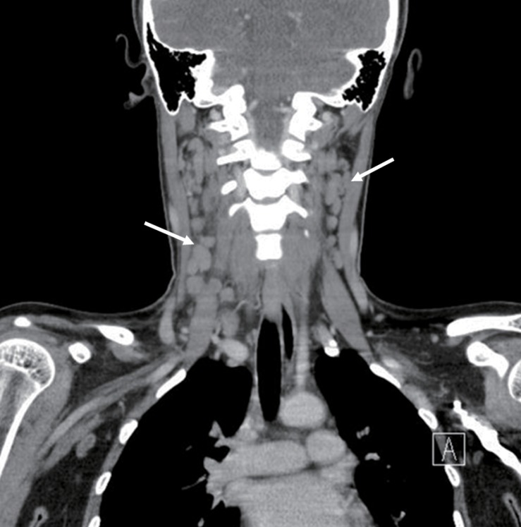
Computed tomography of the neck shows multiple enlarged lymph nodes on both sides of the neck (arrows).
On the second day of hospitalization, she complained of pruritic skin rashes on her lower extremities. Despite antibiotic and analgesic treatment, the fever persisted, the skin rashes spread to her trunk and upper extremities, her cervical lymph nodes continued to enlarge, and the lymphadenopathy spread to the occipital area. On the sixth day of hospitalization, an excisional biopsy of the enlarged cervical lymph node was performed, and the histopathologic findings were consistent with KFD (Fig. 2). Her fever persisted after the excisional biopsy, so we started the administration of oral prednisolone (0.5 mg/kg/day) on the seventh day of hospitalization. On the ninth day of hospitalization, the fever disappeared and the skin rashes began to subside. Ultrasonography of her thyroid showed heterogeneous echogenicity of the thyroid gland and a solitary nodule.

Histopathologic findings of the excised lymph node (H&E, ×400). The excised lymph node shows numerous histiocytes with karyorrhectic debris in the necrotic lesion, and the lesion lacks neutrophils and eosinophils.
We diagnosed her with autoimmune thyroiditis on the basis of her family history and the laboratory results. Thus, she was ultimately diagnosed with KFD and autoimmune thyroiditis. Her hemoglobin was 9.2 g/dL after one month of iron supplementation.
Discussion
KFD was first described in 1972 separately by Kikuchi6) and Fujimoto et al.7). It has a worldwide distribution with a higher prevalence in Southeast Asian women2). KFD usually occurs in young women between the ages of 20 and 40 years1). The female to male ratio is 4:11), but some reports from East Asia show a relatively lower female to male ratio, 1:1 to 2.28:18-10).
KFD commonly manifests as cervical lymphadenitis with fever, but mediastinal, mesenteric, inguinal, or axillary lymph nodes may be involved2,10). Other uncommon symptoms include upper respiratory symptoms, sore throat, weight loss, fatigue, headache, nausea, vomiting, arthralgia, skin rash, and hepatosplenomegaly1,2,9,10). Leucopenia, anemia, elevated ESR, elevated serum LDH, or elevated liver enzyme levels have been reported in many cases, although there is no specific laboratory finding indicative of KFD1,2,8-10).
KFD can be diagnosed definitively through histological analysis of lymph node biopsy tissue1). Fine-needle aspiration cytology (FNAC) can be used to diagnose KFD11). However, the diagnostic accuracy of FNAC for KFD is about 56%, thus excisional biopsy of the involved lymph node is necessary for the accurate diagnosis of KFD1). The involved lymph nodes characteristically demonstrate apoptotic necrosis with abundant karyorrhectic debris in the cortical and paracortial areas1,12). The necrotic lesions are surrounded by many different types of histiocytes including plasmacytoid dendritic cells, and the number of transformed lymphocytes with immunoblast morphology is increased in some cases1,12). However, neutrophils and eosinophils are characteristically absent, and plasma cells are few or absent1,12). Lymphocytes are predominantly T cells rather than B cells, and the T cells are predominantly CD8+ T cells rather than CD4+ T cells1,12).
The cause and pathogenesis of KFD remains unclear, and two hypotheses have been suggested: a viral infection hypothesis and an autoimmune hypothesis1). Prodromal upper respiratory symptoms, the lack of response to antibiotic therapy, atypical lymphocytes in some cases, T cell zone expansion with immunoblast proliferation, and elevated interferon-α support the hypothesis of a viral etiology1). Many viruses including EBV, cytomegalovirus, HHV, parvovirus B19, and HTLV-1 were suggested as the causative organism, but a causal relationship has not been confirmed2). In contrast to the pathologic findings of KFD, viral lymphadenitis has less prominent histiocyte infiltration, more neutrophils and plasma cell proliferation, and CD4+ T cell predominance1).
Based on the clinical similarity (fever, lymphadenitis, skin rash, arthralgia) and histologic similarity (paracortical necrosis with karyorrhectic debris and inflammatory cell responses) of KFD and SLE, an autoimmune etiology was suggested3,12). In addition, continuing reports of autoimmune diseases in KFD patients and therapeutic response to steroids and intravenous immunoglobulins in KFD patients with autoimmune diseases also support the autoimmune origin of KFD3). KFD cases with SLE, HLH, Sjögren syndrome, adult onset Still's disease, and polymyositis have been reported from several areas of the world, and the autoimmune diseases developed before, after, or simultaneously with KFD2-5,10,13).
However, only a few KFD cases with autoimmune thyroiditis have been reported3,14). Until now, a case of KFD with autoimmune thyroiditis has not been reported in Korea. We diagnosed this 17-year-old girl with KFD on the basis of the histopathologic findings of the excised lymph node, and autoimmune thyroiditis was diagnosed on the basis of the presence of antithyroid antibodies. This case is an additional example of the relationship between KFD and autoimmune pathogenesis.
KFD is a self-limiting condition and usually resolves in one to six months with recurrence rates of 3 to 4%1,9). Usually, only symptomatic treatment with analgesics and antipyretics is needed1). Steroids can be used to treat patients with complicated or systemic KFD, recurrent KFD, prolonged fever, and symptoms lasting more than two weeks despite treatment with nonsteroidal antiinflammatory drugs14). In our case, lymphadenopathy spread to the occipital region and the fever remained after the excisional biopsy, so we decided to treat her with oral steroids, and there was a dramatic clinical effect.
To date, several KFD cases associated with various types of autoimmune diseases have been reported4,5,13,15). These reports support the hypothesis that KFD is caused by an autoimmune mechanism. Clinicians should consider KFD when lymphadenopathy arises in patients with autoimmune disease, and should also keep in mind the possibility of autoimmune disease in KFD patients. However, it is not clear whether KFD and autoimmune diseases have a direct association or occur concomitantly by chance2). Additional evaluations of the relationship between KFD and autoimmune diseases are necessary.