All issues > Volume 67(12); 2024
Right ventricular mass in a 10-year-old girl with osteosarcoma: an unusual case of asymptomatic cardiac metastasis
- Corresponding author: Jun Ah Lee, MD. Department of Pediatrics, National Cancer Center, 323 Ilsan-ro, Ilsandong-gu, Goyang 10408, Korea Email: junahlee@ncc.re.kr
- Received May 28, 2024 Revised July 3, 2024 Accepted July 17, 2024
During routine surveillance imaging of a 10-year-old girl with osteosarcoma, a right ventricular filling defect was identified on contrast-enhanced chest computed tomography (CT). Sixteen months earlier, she was diagnosed with osteosarcoma of left distal femur, with lung metastasis. Response to chemotherapy was poor. After limb salvage surgery, chemotherapy was changed, followed by bilateral wedge resection of lungs. However, she developed another lung metastasis one month later. Despite switching chemotherapy, she experienced left buttock pain. Magnetic resonance imaging (MRI) revealed lumbar spine metastasis. Radiation therapy and extradural tumor excision provided temporary pain relief. Chemotherapy was changed, however, her tumor rapidly progressed to involve thoracic spine, and left femur.
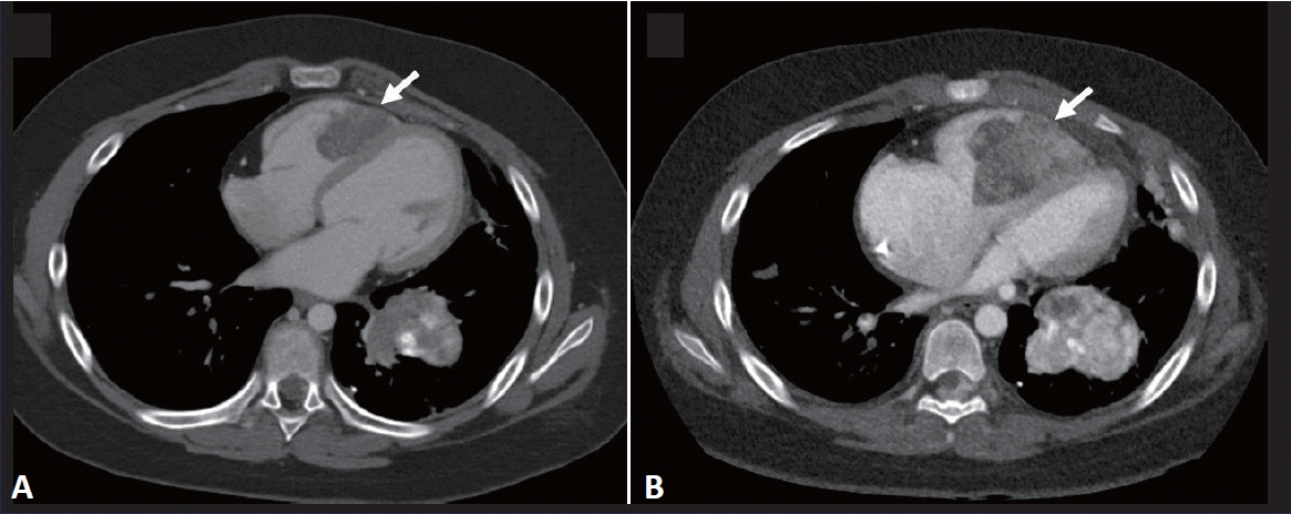
She showed no respiratory or cardiac symptoms, and vital signs were normal. Physical examination revealed no signs of heart murmur, lower limb swelling, or hepatomegaly. Contrast-enhanced chest CT revealed multiple nodules, along with an unexpected filling defect in right ventricle (Fig. 1). The pulmonary artery and inferior vena cava were patent. Echocardiography demonstrated an irregular surfaced mass measuring 4.1 cm×1.8 cm in the right ventricle occupying right ventricular outflow tract (Fig. 2). Right ventricle was slightly dilated with suspicious dysfunction. Trivial tricuspid regurgitation and mild pulmonary hypertension (right ventricular systolic pressure, 40 mmHg) were observed. Left ventricular size and contractility were normal. Laboratory findings were as follows: prothrombin time, 13.6 seconds (international normalized ratio, 1.06); activated partial thromboplastin time 33.4 seconds; fibrinogen, 391 mg/dL (reference rage, 200–400 mg/dL); and D-dimer, 1.85 μg/mL (reference rage, 0–0.39 μg/mL). She had central line (chemoport), however, considering the mass location and her advanced disease status, right ventricular mass was presumed to be a metastatic tumor.
After discussing her grave condition, the parents opted for palliative care. She remained relatively well, however, she complained of chest tightness and pain 6 weeks later. Chest radiography revealed bilateral pleural effusions. She succumbed to progressive respiratory distress 4 days later.
The present study was approved by the Institutional Review Board of National Cancer Center in Korea (NCC2024-0047), which waived the requirement for informed consent because of the retrospective nature of the study.
Our case is a rare presentation of noncontiguous metastasis of osteosarcoma to right ventricle. Cardiac metastasis has been reported in patients with lung, breast, stomach, and liver carcinomas; lymphoma; leukemia; and melanoma [1]. When metastasis occurs, 75% of cases involve pericardium/epicardium, and most present as a pericardial effusion [2]. In children, cardiac metastasis was reported in patients with non-Hodgkin lymphoma, neuroblastoma, Wilms’ tumor, malignant teratoma, and pleuropulmonary blastoma [3]. Few cases in the literature describe cardiac involvement of osteosarcoma, occurring via hematogenous spread or direct extension from pulmonary veins or vena cava [4]. These events are less well-described condition, often incidentally detected during staging investigation.
Few cases are reported with direct cardiac involvement of osteosarcoma [5,6]. Patients with cardiac involvement of osteosarcoma are often asymptomatic and, therefore, can be difficult to recognize [7]. Considering the hematogenous spread of osteosarcoma, frequency of cardiac involvement may have been underestimated, especially in cases of relapsed or refractory tumors. A prevalence as high as 20% has been observed at autopsy of patients with osteosarcoma, suggesting that cardiac involvement is a late stage of hematogenous metastasis [8]. In a series of 20 patients with cardiovascular involvement of osteosarcoma, 3 patients had noncontiguous cardiac tumor involvement, while the other 17 exhibited direct intravascular extension from primary or pulmonary arterial metastatic lesions [4]. Cardiac involvement was more frequent in patients with pelvic than with distal femoral primary lesions, related with the proximity of tumor to large central blood vessels [6]. Meanwhile, cardiac metastasis has been reported in patients with lower extremity osteosarcoma [5,7].
Intracardiac masses in cancer patients present a diagnostic challenge. Due to the invasive nature of biopsy procedures, obtaining pathologic confirmation is often precluded in cancer patients. The enhanced resolution of modern CT holds the potential for improved detection of cardiac involvement. Contrast-enhanced chest CT chest would improve detection of cardiovascular metastases of osteosarcoma [4]. Mineralization is obscured by contrast agent, and nonmineralized tumor thrombi in large vessels and heart may be obscured without contrast agent [4]. Echocardiography is a noninvasive procedure and useful in diagnosis, tumor differentiation and follow-up [2]. Echocardiography has shown greater sensitivity than MRI for detection of intramural and intracavitary tumor, while MRI has increased ability to detect apical tumors [2]. Another complicating factor about cardiac mass in a cancer patient is the differential diagnosis between tumors and bland thrombi. Incidence of bland thrombus or deep venous thrombosis is very low in children younger than 15 years, with less than 5 per 100,000 [9]. And bland thrombi either improve or resolve on follow-up imaging studies; even if the thrombus is persistent, the luminal caliber of the vein involved by a bland thrombus is relatively small [4]. On the other hand, tumor thrombus shows heterogeneous enhancement on contrast-enhanced CT [4].
Management of cardiac involvement in osteosarcoma is often limited to palliative measures, with such involvement typically observed at advanced stages [4]. Usually, patients already have metastasis to axial skeleton, as well as the lungs, after undergoing multimodal treatments. Our patient had previously undergone surgery and received multiple lines of chemotherapy; however, the tumor proved refractory and progressed to involve lungs, spine, and pelvis.
Cardiovascular involvement is a possibility in patients with relapsed or refractory tumors. Regardless of clinical signs or symptoms, careful monitoring of cardiac status is necessary when patients undergo imaging.
- Footnotes
-
Conflicts of interest No potential conflict of interest relevant to this article was reported.
Funding This study received no specific grant from any funding agency in the public, commercial, or not-for-profit sectors.
-
Fig. 1.
(A) Axial contrast-enhanced chest computed tomography (CT) image showing a metastatic mass in the right ventricle (arrow) and pulmonary metastasis with calcification in the left lower lobe. (B) CT image taken 1 month later demonstrating increased mass size in the right ventricle with extension to the tricuspid valve and progression of the left lower lobe metastasis (arrow).

Fig. 2.
Echocardiography showing a tumor mass in the right ventricle. (A) Modified apical 4-chamber view (arrow). (B) Right ventricular inflow tract view. (C) Subcostal view.

- References
- 1. Reynen K, Köckeritz U, Strasser RH. Metastases to the heart. Ann Oncol 2004;15:375–81.
[Article] [PubMed]2. Ragland MM, Tak T. The role of echocardiography in diagnosing space-occupying lesions of the heart. Clin Med Res 2006;4:22–32.
[Article] [PubMed] [PMC]3. Huh J, Noh CI, Kim YW, Choi JY, Yun YS, Shin HY, et al. Secondary cardiac tumor in children. Pediatr Cardiol 1999;20:400–3.
[Article] [PubMed]4. Yedururi S, Morani AC, Gladish GW, Vallabhaneni S, Anderson PM, Hughes D, et al. Cardiovascular involvement by osteosarcoma: an analysis of 20 patients. Pediatr Radiol 2016;46:21–33.
[Article] [PubMed] [PMC]5. Sbai MA, Ben Hmida N, Daas S, Khorbi A, Souissi M, Marzouk R, et al. Cardiac metastasis of a femoral osteosarcoma. Tunis Med 2010;88:860–1.
[PubMed]6. Platonov MA, Turner AR, Mullen JC, Noga M, Welsh RC. Tumour on the tricuspid valve: metastatic osteosarcoma and the heart. Can J Cardiol 2005;21:63–7.
[PubMed]7. Elasfar A, Khalifa A, Alghamdi A, Khalid R, Ibrahim M, Kashour T. Asymptomatic metastatic osteosarcoma to the right ventricle: case report and review of the literature. J Saudi Heart Assoc 2013;25:39–42.
[Article] [PubMed] [PMC]

 About
About Browse articles
 For contributors

