


 ,
,
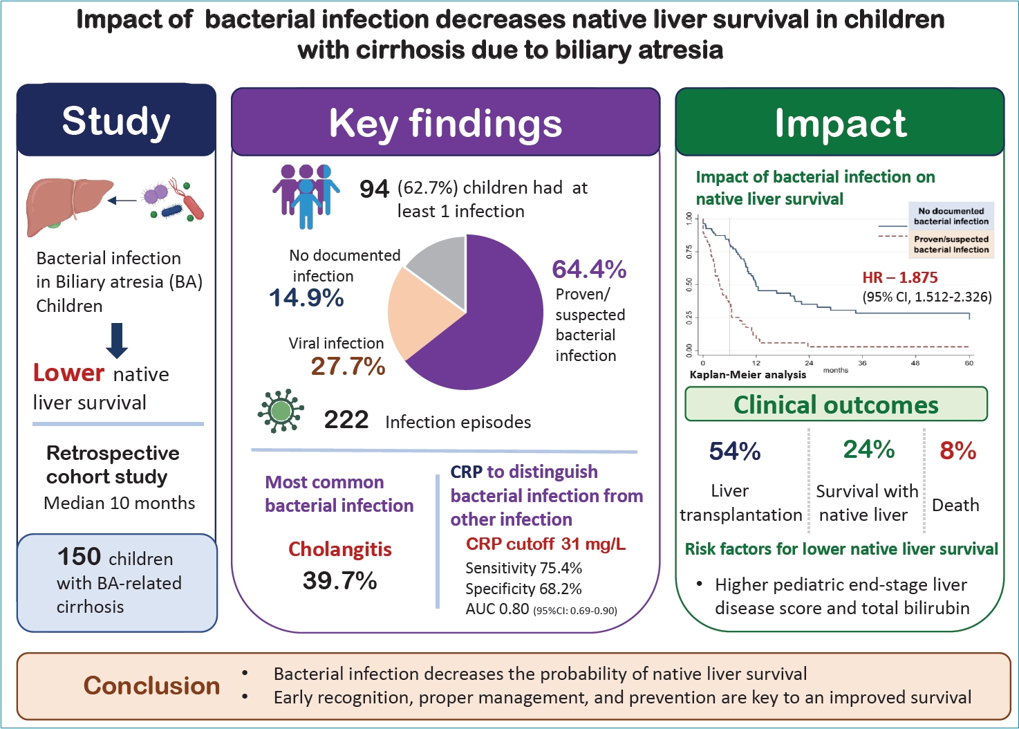
Graphical abstract. CRP, C-reactive protein; AUC, area under the curve; HR, hazard ratio; CI, confidence interval.
Introduction
Cirrhosis is one of the long-term sequelae of biliary atresia (BA) in pediatric patients [1]. Children with BA with delayed diagnosis or could not have adequate bile flow after hepatic portoenterostomy (also known as Kasai operation) generally experience several cirrhosis-related complications such as portal hypertension and variceal bleeding, delayed growth, and malnutrition [2]. Furthermore, a number of children with cirrhosis can also develop potential infection-related life-threatening conditions such as acute cholangitis, spontaneous bacterial peritonitis (SBP), and sepsis/bacteremia [2]. Multifactorial pathogenic mechanisms can exert an excessive inflammatory response and thus induce circulatory dysfunction and acute decompensation that leads to morbidity and death [3].
While there have been numerous studies exploring the characteristics of infections in adult patients with cirrhosis, there is a lack of research in children. The characterization of impact of infections such as septicemia [4,5], SBP [6], acute cholangitis [7], infected biloma in children with BA [8,9], and urinary tract infection have been reported [10]. However, these children can also suffer from other common viral infections such as upper and lower respiratory tract infections, otitis media, and acute gastroenteritis. The incidence of various infections in the tropical countries may be even higher when compared to a report from westernized countries [11]. The evaluation for potential etiologies can therefore be challenging, time-consuming and labor-intensive which can also make the management more difficult or further delayed when compared to the children without cirrhosis. A recent study by Shiau et al. [12] also found that age at Kasai operation and platelet count are risk factors for serious infections.
Moreover, as signs and symptoms can be nonspecific, several biomarkers including C-reactive protein (CRP) [13], procalcitonin [14], or interleukin (IL)-6 [15], have been used to aid and differentiate serious bacterial infection with other less severe infections in patients with cirrhosis; but the yield of the aforementioned markers remain inconclusive [12]. In adults, septic patients with pneumonia or >1 site of infection had a higher mortality [16]. A recent study by Singh et al. found that ascites fluid infection (AFI) was found in 31% children with chronic liver disease and almost half of them were nosocomial infection resulting in high mortality. It was also concluded that children with AFI and worsening cirrhosis should be evaluated for liver transplantation (LT) [17]. Children with cirrhosis who develop several episodes of cholangitis and septicemia are also at risk for LT18) and death [11,12].
With the limited data on this regard in children with BA with cirrhosis, therefore we aimed to study on the characteristics of infection, evaluation and management, potential risk factors for developing infection, clinical outcomes, which remain a gap in the current knowledge. Such studies would likely provide benefits for patient care in effectively managing BA children presenting with suspected infection and possibly leading to an improved prognosis.
Methods
1. Study design, population, and data collection
A single-center retrospective cohort study was performed at the Faculty of Medicine Ramathibodi Hospital, Bangkok, Thailand, a tertiary care teaching hospital with an active LT program for children. The study protocol was approved by the Human Research Ethics Committee of the Faculty of Medicine Ramathibodi Hospital, Mahidol University (approval no. MURA2023/951). The requirement for informed consent was waived because of the retrospective nature of the study. Data collection was performed via electronic medical records and hospital database (International Classification of Diseases, 10th Revision [ICD-10] of Q442) for a period of 10 years (January 2014 to December 2023) in patients age less than 18 years who were diagnosed with cirrhosis due to BA. Patients were excluded if a significant portion of clinical data was missing (e.g., missing primary outcome, missing infection status). Infection episodes were excluded if they were classified as hospital-acquired infections.
According to our objective, we primarily aimed to study pre-LT related infections in patients with cirrhosis due to BA when they presented with fever in outpatient or emergency department. Data included demographic information, underlying condition including severity and complications of cirrhosis, other medical conditions such as acute kidney injury and variceal bleeding. Significant portal hypertension was defined if the patient had evidence of varices, ascites, or thrombocytopenia with splenomegaly [18]. Data on infections including types of infection, diagnosis, clinical courses, laboratory investigations (e.g., CRP, procalcitonin) and treatment. Clinical outcomes were recorded included with LT, survival with native liver, death, or loss to follow-up. These data were retrieved since the diagnosis of BA until either the point of predefined clinical outcome or the last day of followup. Other interested outcomes were the recurrence of infections, worsening liver synthetic function, and complications of cirrhosis such as portal hypertension or variceal bleeding.
2. Definition of proven/suspected bacterial infections
Various proven/suspected bacterial infections would be categorized as a clinically compatible infectious syndrome diagnosed by the treating physician and/or supported by positive-microbiological confirmation into one of the following: Hepatobiliary infection including; acute cholangitis was defined by the Tokyo guideline 2018 criteria as suspected diagnosis and definite diagnosis [19]. Infected biloma was defined by fever with/without increasing jaundice and the evidence of biloma by imaging (liver ultrasonography or computed tomography of the abdomen) [20]. AFI; SBP was defined by polymorphonuclear cells >250 mm3 in the ascitic fluid with positive culture, culture-negative neutrocytic ascites was defined by polymorphonuclear cells >250 mm3 with negative ascitic culture in the absence of previous antibiotic administration, and monomicrobial non-neutrocytic bacterascites was defined by polymorphonuclear cells <250 mm3 with positive ascitic fluid culture [21]. ‘Bacteremia/septicemia’ was defined by fever with positive blood culture [22]. Infectious diarrhea was defined by diarrhea with positive stool culture for enteropathogenic organisms or pathogenic bacteria detected by polymerase chain reaction-based stool tests [23]. Urinary tract infection was defined by fever, pyuria with white blood cells ≥5/high power field or positive leukocyte esterase and/or positive nitrite with positive urine culture [24]. The aforementioned bacterial infections were common in BA patients based on the previous study [12].
3. Definition of presumed viral infections
Presumed viral infection was defined as febrile manifestation and negative for bacterial culture or strong evidence for suspected bacterial infection (e.g., acute cholangitis) with or without positive viral antigen or polymerase chain reaction [12]. Of note, one fever episode could have >1 positive test for either bacteria or viral infection or both, and if they had at least 1 diagnosis of proven/suspected bacterial infection, that fever episode was categorized as bacterial infection.
4. Biostatistical analysis
We performed descriptive statistics to summarize the characteristics, incidence, and types of infection of the study population. Statistical analyses were conducted by using chi-square test or student t test to identify significant associations between risk factors and infections. The Cox proportion hazard model with multiple record and multiple events (applying the covariates, fixed effect) was performed for comparing survival rate between proven/suspected bacterial infection versus presumed viral infection to LT and death.
Results
1. Baseline characteristics
Patients were identified from the hospital database using the ICD-10 code Q442 over a 10-year period, yielding a total of 201 cases, and 50 patients were excluded (Supplementary Fig. 1). Therefore, the total number of patients were 150 patients (females 51.3%, underwent Kasai operation 72%, mean age of the first visit to our center of 6 months, and decompensated cirrhosis in 77.2%). Patients can be categorized into 3 infection-related groups: proven or suspected bacterial infection (n=65), presumed viral infection (n=29), and no documented infection (n=56) as shown in Table 1. The median follow-up duration was 10.2 months to clinical outcomes including LT, survival with native liver, death, and loss to follow-up. No statistically significant differences of each variable were observed among groups with regards to demographics and baseline clinical condition/severity.
2. Infection episodes and diagnosis
A total of 222 fever episodes resulted in 249 infection-related diagnoses, with 64.4% (N=143) classified as proven or suspected bacterial infections and 35.6% (N=79) categorized as presumed viral infections (Table 2). Twenty-seven fever episodes had >1 infection-related diagnosis. Among bacterial infections, the most common diagnoses were acute cholangitis, followed by bacteremia/septicemia, infected biloma, and AFI. Other notable bacterial infections included infectious diarrhea, urinary tract infection, and liver abscesses. Presumed viral infections included gastroenteritis, respiratory tract infections, and others such as exanthem, herpangina.
3. Factors associated with proven or suspected bacterial infection
Four factors were associated with cases with proven or suspected bacterial infection: pediatric end-stage liver disease (PELD) score at the first visit, PELD score at the fever episode, total bilirubin, and direct bilirubin levels during the fever episode (Table 3). In the multivariate analysis, after adjusting for potential confounders, only total bilirubin at the fever episode remained a significant predictor of bacterial infection (hazard ratio [HR] 1.73, 95% confidence interval [CI], 1.09–2.76, P=0.021). However, due to significantly high collinearity with PELD score and bilirubin levels, these variables could not be included in the multivariate analysis.
4. Factors associated proven or suspected bacterial infection (fever episodes)
In the univariate analysis, several factors were found to be associated with episodes of proven or suspected bacterial infection including anemia (defined by hemoglobin <11 g/dL), elevated CRP and PELD score at the time of fever episode. In the multivariate analysis, 2 factors remained an independent predictor: CRP and PELD score (Table 4). Other variables such as high-grade fever, change of mental status, shock, abnormal white blood cell counts (leukocytosis or leukopenia), thrombocytopenia, elevated procalcitonin, and hypoalbuminemia were not statistically significant.
5. CRP to distinguish proven/suspected bacterial infection from presumed viral infection
To be able to differentiate bacterial versus viral infection at the fever episode, CRP as the only significant independent predictor among the biomarkers in the regression analysis was applied to quantify the impact of variability among individuals’ decision thresholds. The receiver operating characteristic (ROC) curve showed a good diagnostic performance of CRP with an optimal cutoff of 31 mg/L, yielding an area under the ROC curve of 0.80 (95% CI, 0.69–0.90) as shown in Supplementary Fig. 2. At this cutoff, CRP demonstrated a sensitivity of 75.4% and specificity of 68.2%. The positive predictive value and negative predictive value were 87.5% and 93.8%, respectively, with an overall accuracy of 73%.
6. Independent factors contributing to the chance of undergoing LT
Table 5 presents univariate and multivariate Cox regression analyses to identify factors associated with LT. In the univariate analysis, proven or suspected bacterial infection, and elevated PELD score at the first visit to our center and last visit were both associated with a higher chance of LT. However, only proven or suspected bacterial infection remained an independent predictor of LT. Not only for LT, but mortality also affected by bacterial infection with an adjusted HR of 51.03 (P<0.001).
7. Impact of bacterial infection on native liver survival
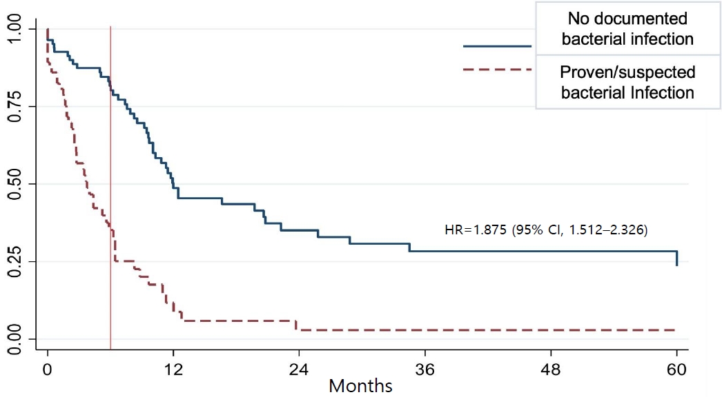
Patients with proven or suspected bacterial infection had significantly poorer native liver survival (i.e., more LT or death) compared to those without documented bacterial infection. The survival probability in the bacterial infection group (dashed red line) declined rapidly, particularly within the first 6 months after the first visit (vertical red line), whereas the nonbacterial infected group (solid blue line) demonstrated a more sustained native liver function over time. The probability of native liver survival dropped to approximately 25% at 6 months in children previously suffered from bacterial infection while the ones without bacterial infection had a chance of maintaining a native liver status at 80%. The HR for loss of native liver in the presence of bacterial infection was 1.88 (95% CI, 1.51–2.33), indicating a nearly twofold increased risk (Fig. 1).
The clinical factors associated with outcomes in children with BA, categorized into 4 groups: LT (n=81), survived with native liver (n=36), death (n=12), and loss to follow-up (n=21). A significantly higher proportion of patients in the native liver group were the ones who underwent Kasai operation (94.4%) was noted, when compared to the LT group (65.4%) (P=0.008), similarly to as the presence of decompensated cirrhosis (P<0.001) (Table 6). PELD score at the first visit also varied significantly across the 4 groups (P<0.001). Other variables, such as sex, age at Kasai operation after 90 days, and history of past infection before referral to our center did not show statistically significant differences among groups.
Discussion
In a single-center cohort of BA children with cirrhosis, bacterial infections were common, with cholangitis being the most common bacterial infection, accounting for nearly 40% of total bacterial infection. Other liver disease-related infections such as infected biloma and AFI were also relatively common in our population. While comparing between the 3 groups (bacterial infection, viral infection, and no infection), no statistically significant differences of demographics, performed Kasai operation, or baseline clinical condition were observed. This finding is consistent with a study from Taiwan [12] that reported cholangitis in 46.2% of post-Kasai patients, reinforcing its role as a leading infectious complication in this population.
Bacterial infections were associated with significantly higher PELD scores both at the first visit and the fever episode as well as bilirubin levels as a person-related data (Table 3). These parameters reflect the severity of liver dysfunction in these children with chronic liver disease. Similarly, Bolia et al. [25] demonstrated that a higher Child-Pugh score, along with lower serum albumin and elevated international normalized ratio, was associated with an increased risk of infection in children with acute liver failure and decompensated chronic liver disease. This finding was parallel between Child-Pugh and PELD scores across different studies emphasizing the relevance of liver disease severity in predisposing to various types of infections. Cirrhosis is an immunomodulatory deficiency state [26], predisposing such patients to other various infections through multiple mechanisms [19,27], such as bacterial overgrowth and small intestinal barrier dysfunction resulting in further bacterial translocation [4,6,27,28]. Genetic variants in those with cirrhosis have also been noted to be associated with an increased risk and severity of infection [29].
From Table 4 as described the infection episode-related data, CRP and PELD score were 2 significant independent associated factors of proven/suspected bacterial infection (vs. presumed viral infection). Other clinical and laboratory parameters such as high-grade fever, abnormal blood cell indices, procalcitonin or hypoalbuminemia were not statistically significant. A recent study by Shiau et al. [12] also found that platelet count is one of the risk factors for future serious infections. From our study, CRP levels showed good discriminatory power with an area under the ROC curve of 0.80 at a cutoff of 31 mg/L, yielding an accuracy of 73%. This finding may support the clinical utility of CRP as a biomarker for an identification of bacterial infection in this population. However, this cutoff value is notably higher than those reported in previous pediatric studies. For instance, Bolia et al. [30] and Kalvandi et al. [31] reported an optimal threshold of 6 mg/L in children with acute liver failure, decompensated chronic liver disease, or ascites, with good diagnostic performance for bacterial infections and mainly for SBP. Similarly, Deutsch et al. [13] demonstrated that a CRP cutoff of 10 mg/L was appropriate for detecting bacterial infections in adult patients with cirrhosis. This relatively high CRP threshold and limited number of documented test maybe due to that CRP was being performed selectively in cases with more severe clinical presentations and/or when followup of treatment response was deemed necessary by the treating physician.
We found that native liver survival was significantly reduced in patients with bacterial infection (adjusted HR, 1.88), reinforcing the role of infection in liver function deterioration and loss of transplant-free survival. Although a higher PELD score was associated with an increased risk of death and transplantation in the univariate analysis, it did not retain statistical significance in the multivariable analysis (Table 5). These findings highlight the detrimental effect of bacterial infection on the preservation of native liver function and suggest that the management of bacterial infection may likely be crucial in improving long-term liver outcomes [27]. Based on our results and existing evidence, cholangitis is the most common cause of proven/suspected bacterial infection in children with BA, likely related to previous Kasai portoenterostomy. Moreover, our study demonstrated that, after adjusting for disease severity scores and prior history of infection before referral, proven/suspected bacterial infection was associated with a lower probability of native liver survival (i.e., higher rates of LT or death). However, there is currently dearth of studies evaluating the clinical outcomes in patients with BA and cirrhosis after bacterial infections. Therefore, this aspect reported in the present study addresses the existing knowledge gap.
This study examined infections in a relatively large cohort of children with BA, encompassing not only bacterial infections but also viral infectious etiologies. We evaluated the utility of clinical and laboratory parameters in differentiating bacterial infections from other infections, and to explore the clinical outcomes associated with bacterial infections. However, several limitations should be acknowledged. First, the retrospective design inherently limits data completeness, and missing data were unavoidable. In addition, data collection was limited to the ‘documented’ infection episodes, resulting in inconsistent timing across patients and preventing longitudinal assessment of changes in clinical status or risk factors over time. Accordingly, this may have contributed to the relatively high proportion of patients classified into the “no documented infection” group. The CRP was measured in just 40% of the total infection episodes as it may be due to that CRP was performed based on clinical indication and selectively checked in cases with more severe clinical presentations or those mostly diagnosed with proven/suspected bacterial infections. Patients with milder clinical presentations, such as low-grade fever, viral illness, or upper respiratory tract infections, were less likely to undergo CRP testing. Therefore, we acknowledge this as a limitation in this retrospective study. The median follow-up duration was approximately 10 months, which may be considered relatively short. This is largely attributable to the fact that most patients in this cohort were referred for LT, and the evaluation and treatment process was typically completed within a relatively short timeframe.
In conclusion, infections mainly cholangitis and bacteremia are common in children with cirrhosis due to BA. A higher PELD score at the first visit and during febrile episodes, as well as elevated bilirubin levels, were significantly associated with proven or suspected bacterial infection. Specific clinical and laboratory parameters, such as CRP, are useful in differentiating bacterial infections from other causes of fever. Bacterial infection was also associated with a decreased early survival rate. Developing appropriate strategies to manage and prevent such bacterial infections could improve clinical outcomes.


 PDF Links
 PubReader
 ePub Link
 XML Download
 PubMed
 Download Citation