


 ,
, Graphical abstract
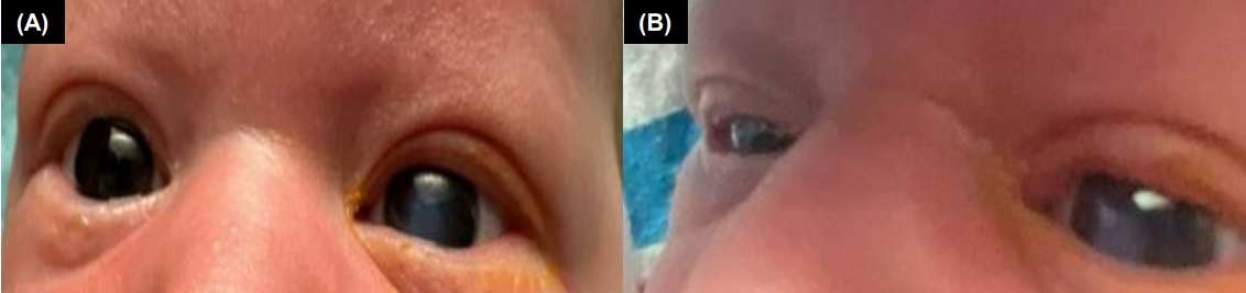
A 1-day-old male, born full-term via normal spontaneous vaginal delivery to a healthy mother, is seen for routine care. Prenatal ultrasound at 32 and 37 weeks’ gestation showed right sided grades 2–3 (8.9 mm and 9.7 mm, respectively) urinary tract dilation (UTD). Delivery was uneventful and atraumatic. On examination, left eye corneal clouding (Fig. 1A and B) and preauricular pits bilaterally were noted. The red reflex could not be visualized on the left eye. The right eye, including the red reflex, was normal. The remainder of the exam was benign. Family history revealed no eye or genetic abnormalities.
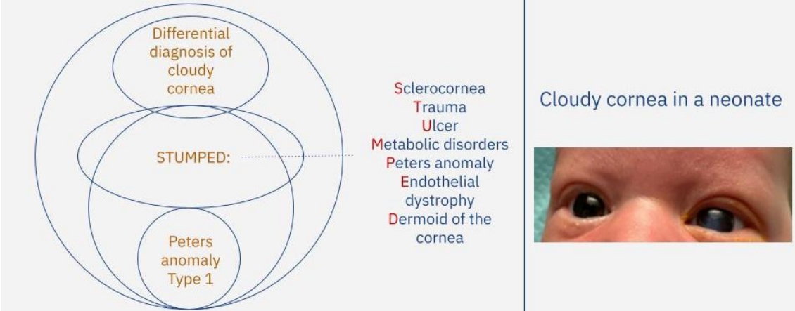
The differentials for corneal clouding include Descemet’s membrane tears, metabolic disorders, corneal edema, cataracts, glaucoma, birth trauma, rubella, herpes simplex virus keratitis, and Axenfeld-Reiger Syndrome. Sclerocornea, Peters anomaly (PA) and limbal dermoid are rare and should be considered. The “STUMPED” mnemonic: Sclerocornea, Trauma, Ulcer, Metabolic disorders, PA, Endothelial dystrophy, and Dermoid of the cornea, aids in the differential of cloudy cornea [1].
Pediatric ophthalmology evaluated the patient on day of life (DoL) 1. Left eye evaluation revealed a dense, central opacity of the cornea with no fluorescein uptake. Red reflex was not visualized. The posterior pole was poorly visualized, with adhesions from iris to cornea, without lens involvement. Eye palpation was soft, suggesting no increase of intraocular pressure. The right eye exam was normal. The presumptive diagnosis was PA, a defect in the corneal posterior stroma, Descemet’s membrane, and endothelium, resulting in a corneal opacity.
He was discharged on DoL 3 after routine care with close outpatient ophthalmology follow-up and referral to a transplant specialist. Interim serial eye pressure measurements were normal. Four months after discharge, the patient had successful penetrating keratoplasty (PK). Surgical pathology did not show lens involvement, confirming the diagnosis of PA type 1. Although Peters-Plus was initially considered due to the associated UTD, a subsequent renal bladder ultrasound showed resolved UTD, making this unlikely. The infant continues to do well on serial follow-up visits.
PA results from aberrant migration of neural crest cells and is characterized by opacification of the cornea, developmental glaucoma, subcapsular cataract, and thinning of Descemet's membrane [2]. Glaucoma is present in more than 50% of the cases [2,3]. It is rare, with an incidence of 1–1.5 per 70,000–100,000 [3,4]. Inheritance patterns may be autosomal dominant, recessive, or sporadic. The majority of cases are bilateral, as unilateral presentations are typically an isolated finding [1]. Common genetic mutations include CYP1B1 (autosomal recessive), and PAX6, PITX2, and FOXC1 (autosomal dominant). Other ocular disorders related to abnormal neural crest cell migration include Axenfeld-Rieger Syndrome (posterior embryotoxon and iris hypoplasia causing pupil displacement), primary congenital glaucoma (abnormal iridocorneal angle and trabecular meshwork formation), and congenital ocular coloboma (fissure closure failure) [5].
PA can be part of fetal alcohol syndrome, congenital rubella, or associated with WAGR syndrome (Wilm tumor, aniridia, genitourinary abnormalities and intellectual retardation), Rubinstein-Taybi syndrome, and PHACES (posterior fossa malformations, hemangiomas, arterial anomalies, cardiac defects, eye abnormalities, sternal cleft, and supraumbilical raphe syndrome) [4].
Three types of PA with diagnostic criteria are listed [6]: type 1: central corneal opacity with iris adhesions without lens involvement; type 2: central opacity with cataracts or lens adhesions; can be associated with microphthalmia, iris coloboma, and retinal abnormalities; type 3 (Peters-Plus): corneal clouding with other associated defects, including short stature and shortened limbs, cleft palate, facial anomalies, intellectual disability, and less commonly heart defects and genitourinary anomalies.
Diagnosis can be made by ophthalmoscopy and confirmed by advanced imaging such as ultrasound biomicroscopy [2]. Genetic confirmation is not required for diagnosis, but may be helpful for family members.
Observation can be considered for small opacities as they may improve spontaneously, but most patients are considered for surgical procedures, as delaying increases the risk of amblyopia and glaucoma [2]. Surgical management includes PK, also known as corneal transplant, or peripheral iridectomy. Potential surgical complications include graft failures, postoperative cataracts, glaucoma, and amblyopia [1,7]. The transplant survival rate is between 50%–70% during the 1st year, but decreases to 30% after 5 years. Intraocular pressure control may require multiple surgeries including placing glaucoma-draining devices [6]. The most beneficial time to perform PK is controversial with poor outcomes seen when performed in very early infancy, while higher complications noted if performed after 2 years of age. The ideal timing for PK appears to be around 5 months of age with the most promising results [2]. Novel treatments such as optical iridectomy and selective endothelial removal are being evaluated [3,8,9].
This case highlights the importance of recognizing newborn examination findings needing urgent ophthalmological evaluation. Cloudy cornea has a wide differential, with PA being uncommon. PA requires referral for early surgical management to prevent complications and frequent postoperative monitoring to evaluate transplant success. For suspected Peters-Plus, a genetics referral is recommended.
Written informed consent was obtained from the parents to use clinical images and details of the child for educational and publication purposes. The institutional review board (IRB) of Jersey Shore University Medical Center determined that this case report was exempt from IRB review.


 PDF Links
 PubReader
 ePub Link
 XML Download
 PubMed
 Download Citation