Article Contents
| Korean J Pediatr > Volume 58(11); 2015 |
|
Abstract
Diphyllobothrium latum infection in humans is not common in Republic of Korea. We report a case of fish tapeworm infection in a 10-year-old boy after ingestion of raw perch about 8 months ago. The patient complained of recurrent abdominal pain and watery diarrhea. A tapeworm, 85 cm in length, without scolex and neck, was spontaneously discharged in the feces of the patient. The patient was treated with 15-mg/kg single dose praziquantel, and follow-up stool examination was negative after one month. There was no evidence of relapse during the next six months.
Diphyllobothrium latum is one of the longest intestinal tapeworms of mammals, such as humans, dogs, cats, foxes, and other wild canines1). The infection by D. latum is common in regions with cold water lakes, such as North America and Europe. In Asia, this infection has been reported in Republic of Korea, Japan, Siberia and Malaysia2). In Republic of Korea, the first case of D. latum infection was reported in Jinju by the recovery of eggs in the feces of 2 patients3). An adult tapeworm was reported for the first time in 19714). A total of 48 cases have been diagnosed by the adult worm from 1971 to 20125). We report here a case of a young child with D. latum infection in Republic of Korea.
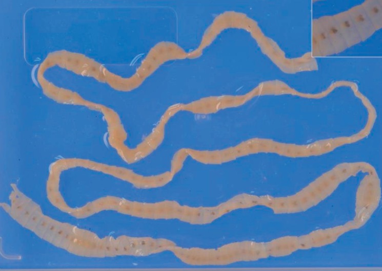
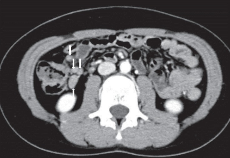
A 10-year-old boy visited our outpatient clinic with a segment of a tapeworm which was naturally discharged in his feces on the previous day. He was a healthy elementary school student in the city, without any recent history of travel to a foreign country. His family lives in urban area (Iksan city, Jeollabuk-do) but his parents reported frequent consumption of raw fish, and ingestion of raw perch about 8 months ago. He had suffered from recurrent lower abdominal pain and watery diarrhea for about 6 months. These episodes were characterized by vague abdominal pain that may be dull or crampy relatively. The abdominal pain frequently relieved with watery diarrhea but relapsed every 2-3 days. And three months ago, he detected several noodle-like whitish materials in his feces. But, he did not visit any medical institution at that time. On physical examination, no specific signs, such as pale conjunctiva and abdominal distention or tenderness were observed. Hematologic examination revealed eosinophilia without signs of anemia and his erythrocyte sedimentation rate was elevated (hemoglobin 13.2 g/dL, hematocrit 37.5%, eosinophil 710/µL, erythrocyte sedimentation rate 29 mm/hr). Blood chemistry and serology were within the normal range. The coprological study was positive for D. latum eggs. The worm was white, 85 cm long without scolex, and was identified as D. latum, based on the biological characters (Fig. 1). To determine the accompanying complications such as intestinal obstruction or cholangitis, we performed radiological examination. Abdominal ultrasonography showed mild intestinal wall swelling and normal hepatobiliary findings. On abdominal computed tomography, multiple mesenteric lymph nodes were enlarged, but no other complications were found (Fig. 2). He was treated with a single oral dose of praziquantel (15 mg/kg), and stool examination was negative one week later. His symptoms improved, and there was no evidence of relapse during the next 6 months.
Diphyllobothriasis, an infection by Diphyllobothrium species, is a zoonosis acquired by humans and other mammals, having a worldwide distribution1). Several species of Diphyllobothrium have been reported in humans. D. latum, Diphyllobothrium pacificum, and Diphyllobothrium nihonkaiense are the main pathogens of human Diphyllobothriasis. D. pacificum is common in South America and D. nihonkaiense is common in Japan. D. latum is almost worldwide in distribution, occurring in northern temperate and subtropical areas of the world6). Creamy white in color, D. latum is one of the longest tapeworms causing human Diphyllobothriasis, measuring 4 to 15 m in length and 10 to 20 mm in width, and may consist of 3,000 to 4,000 proglottids. The scolex is shaped like a spoon and has a pair of bothria, in its anterior portion that serve as an organ of attachment7,8). The eggs of D. latum hatch into coracidium in cool fresh water. These are ingested by copepods (the first intermediate host), and develop into the procercoids. Second intermediate hosts include freshwater, anadromous, or marine fish. Through the ingestion of infected copepods, the procercoids enter their tissues and develop into the plerocercoid stage. Raw fish consumption by a definitive host allows the plercocercoids to attach to the small intestine wall. There, they develop rapidly into a mature tapeworm. Mature tapeworm can live for many years in the host intestine, and can discharge very large numbers of eggs per day, completing the cycle9). D. latum is transmitted to humans through eating raw or uncooked fish. As to the second intermediate hosts for D. latum, freshwater fishes, such as pikes, trouts, salmons, and perches have been reported1). In a study performed in Republic of Korea, salmon, mullet, perch, and trout were reported as the suspected sources of infection10). Our patient also had a history of eating raw fish frequently, and he had consumed raw perch about 8 months ago. The recent rapid advancement of life quality with improvement of dietary conditions in Republic of Korea, in particular, increased consumption of expensive raw fish, is suggested to be a factor responsible for an increase in D. latum infections. Because D. latum infection is caused due to this eating habit, it very rarely occurs in children. Although this infection is rare in children, in Republic of Korea, about 5 cases including the present case have been reported in children up to the age of 10 years (Table 1)10,11). D. latum infections are often asymptomatic. Clinical manifestations are often mild and vague, including fatigue, constipation, and poorly defined abdominal discomfort. Other symptoms of this disease are pernicious anemia, headache, and allergic reactions9). The symptoms reported by the patient in this study were recurrent abdominal pain and watery diarrhea. These were similar to those in a previous study9). Laboratory investigations tend to present normal results, but in prolonged or heavy D. latum infection, they might show low vitamin B12 levels or frank pernicious anemia12,13). Only history taking and considering the possibility of parasitic infection can avoid further invasive evaluation. Praziquantel is used as the drug of choice in D. latum infection, and it is usually given as a single dose of 10 to 25 mg/kg. From 1987 up to present, praziquantel had been used primarily, and there has been no treatment failure among the 43 cases reported in Republic of Korea10). In our case, a 15 mg/kg single dose of praziquantel was used. One week later and one month later, the patient underwent stool examination for D. latum, and it was negative. Niclosamide and intraduodenal gastrografin have been reported to be efficacious as an alternative therapy for diphyllobothriasis9). Because cases of D. latum infection in Republic of Korea are expected to continue to increase and the symptoms are vague, physicians should give more consideration to cases in children with recurrent abdominal symptoms of unknown cause. Because gastrointestinal fiberoptic endoscopy, upper and lower gastrointestinal series and abdominal magnetic resonance imaging, etc. in an attempt to establish a tentative diagnosis are expensive and painful, it is important that diagnosis of these children with recurrent abdominal pain should begin with a detailed history such as dietary or bowel habit.
Conflicts of interest
Conflict of interest:
No potential conflict of interest relevant to this article was reported.
References
1. Beaver PC, Jung RC, Cupp EW. Clinical parasitology. 9th ed. Philadelphia: Lea & Febiger, 1984.
2. Chou HF, Yen CM, Liang WC, Jong YJ. Diphyllobothriasis latum: the first child case report in Taiwan. Kaohsiung J Med Sci 2006;22:346–351.


3. Kojima R, Ko T. Researches on intestinal parasites of Koreans in South Keisho-Do: especially on the distribution of liver flukes. J Chosun Med Assoc 1919;26:42–86.
4. Cho SY, Seo BS, Ahn JH. One case report of Diphyllobothrium latum infection in Korea. Seoul J Med 1971;12:157–163.
5. Choi HJ, Lee J, Yang HJ. Four human cases of Diphyllobothrium latum infection. Korean J Parasitol 2012;50:143–146.


6. Dick TA, Nelson PA, Choudhury A. Diphyllobothriasis: update on human cases, foci, patterns and sources of human infections and future considerations. Southeast Asian J Trop Med Public Health 2001;32(Suppl 2): 59–76.

7. Lee KW, Suhk HC, Pai KS, Shin HJ, Jung SY, Han ET, et al. Diphyllobothrium latum infection after eating domestic salmon flesh. Korean J Parasitol 2001;39:319–321.


8. Sun T. Diphyllobothriasis, hymenolepiasis, and dipylidiasis. Sun T, editor. Color atlas and textbook of diagnostic parasitology. New York: Igaku-Shoin, 1988;:283–285.
9. Scholz T, Garcia HH, Kuchta R, Wicht B. Update on the human broad tapeworm (genus Diphyllobothrium), including clinical relevance. Clin Microbiol Rev 2009;22:146–160.


10. Lee EB, Song JH, Park NS, Kang BK, Lee HS, Han YJ, et al. A case of Diphyllobothrium latum infection with a brief review of diphyllobothriasis in the Republic of Korea. Korean J Parasitol 2007;45:219–223.


11. Lee JS, Kim BN, Shon YM, Lee JW, Im KI. A case of Diphyllobothrium latum Infection in childhood. Korean J Pediatr Infect Dis 2001;8:129–133.

Fig. 1
A complete strobila without scolex and neck of Diphyllobothrium latum parvum type recovered from the patient. Its whole length was only 85 cm.

Table 1
Summary of five Diphyllobothrium latum cases up to the age of 10 years reported in Korea
| Case No | Study | Sex | Age (yr) | Residence | Complaints | Suspected source | Treatment |
|---|---|---|---|---|---|---|---|
| 1 | Lee et al. (1983)* | Boy | 10 | Seoul | Abdominal pain | Perch | Bithionol |
| 2 | Joo et al. (1983)* | Girl | 6 | Seoul | Anemia | Sea fishes | Bithionol |
| 3 | Joo et al. (1983)* | Boy | 10 | Seoul | Passage of proglottids | Unknown | Bithionol |
| 4 | Lee et al. (2001)* | Girl | 7 | Sokcho | Abdominal pain, | Salmon | Praziquantel |
| 5 | Lee et al. (2001)11) | Girl | 5 | Seoul | Abdominal pain, | Perch | Praziquantel |


 PDF Links
 PubReader
 PubMed
 Download Citation


