Search

  • HOME
  • Search
Review Article
Neonatology (Perinatology)
Quantifying myelin in neonates using magnetic resonance imaging: a systematic literature review
Nabila Hanem Arshad, Hasyma Abu Hassan, Nur Farhayu Omar, Zurina Zainudin
Clin Exp Pediatr. 2024;67(8):371-385.   Published online December 6, 2023
Question: This systematic review attempts to discover the best magnetic resonance imaging (MRI) technique for myelin quantification in neonates by evaluating various MRI parameters and their reproducibility.
Finding: Since the benefits of using synthetic MRI for quantifying myelin in neonates outweigh the very minor draw- backs, it is recommended.
Meaning: The findings suggest the importance of identifying noninvasive MRI techniques available to assess myelin tissue in neonates, which aid in diagnosing neurodevelopmental disorders.
Neurology
Cognitive outcomes in late childhood and adolescence of neonatal hypoxic-ischemic encephalopathy
Bo Lyun Lee, Hannah C. Glass
Clin Exp Pediatr. 2021;64(12):608-618.   Published online May 24, 2021
∙ Cognitive impairments occur in children with hypoxic-ischemic encephalopathy (HIE) even without neuromotor deficits.
∙ Therapeutic hypothermia has improved neurodevelopmental outcomes of children with HIE; however, 40% of children remain at risk of death/disability or cognitive impairments necessitating the development of adjunctive neuroprotective therapies.
∙ Long-term follow-up until adolescence is required to identify cognitive dysfunction.
∙ A pattern of watershed injury on brain imaging is associated with poor cognitive outcomes.
Advanced neuroimaging techniques for evaluating pediatric epilepsy
Yun Jeong Lee
Clin Exp Pediatr. 2020;63(3):88-95.   Published online February 6, 2020
Accurate localization of the seizure onset zone is important for better seizure outcomes and preventing deficits following epilepsy surgery. Recent advances in neuroimaging techniques have increased our understanding of the underlying etiology and improved our ability to noninvasively identify the seizure onset zone. Using epilepsy-specific magnetic resonance imaging (MRI) protocols, structural MRI allows better detection of the seizure onset zone,...
Original Article
Neurology
Correlation of serum S100B levels with brain magnetic resonance imaging abnormalities in children with status epilepticus
Prastiya Indra Gunawan, Darto Saharso, Dian Purnama Sari
Clin Exp Pediatr. 2019;62(7):281-285.   Published online May 8, 2019

Purpose: To evaluate the association between elevated S100B levels with brain tissue damage seen in abnormalities of head magnetic resonance imaging (MRI; diffusion tensor imaging [DTI] sequence) in patients with status epilepticus (SE). Methods: An analytical observational study was conducted in children hospitalized at Dr Soetomo Hospital, Surabaya, from July to December 2016. The patients were divided into 2 groups: SE...
Respiratory syncytial virus-associated seizures in Korean children, 2011–2016
Teahyen Cha, Young Jin Choi, Jae-Won Oh, Chang-Ryul Kim, Dong Woo Park, In Joon Seol, Jin-Hwa Moon
Clin Exp Pediatr. 2019;62(4):131-137.   Published online October 23, 2018

Purpose: Respiratory syncytial virus (RSV) infection can cause various neurological complications. This study aimed to investigate the RSV-associated neurologic manifestations that present with seizures. Methods: We retrospectively reviewed the medical records of patients aged less than 15 years with laboratory-confirmed RSV infections and seizures between January 2011 and December 2016 in a regional hospital in South Korea. Results: During this period, 1,193...
Neonatology (Perinatology)
Patterns of ischemic injury on brain images in neonatal group B Streptococcal meningitis
Seo Yeol Choi, Jong-Wan Kim, Ji Won Ko, Young Seok Lee, Young Pyo Chang
Clin Exp Pediatr. 2018;61(8):245-252.   Published online August 15, 2018

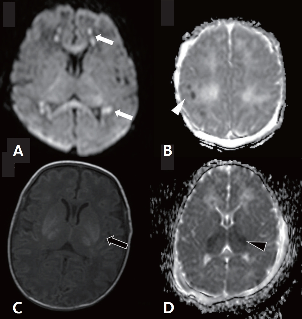
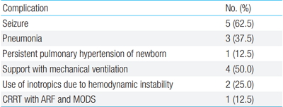
Purpose: This study investigated patterns of ischemic injury observed in brain images from patients with neonatal group B Streptococcal (GBS) meningitis. Methods: Clinical findings and brain images from eight term or near-term newborn infants with GBS meningitis were reviewed. Results: GBS meningitis was confirmed in all 8 infants via cerebrospinal fluid (CSF) analysis, and patients tested positive for GBS in both blood and CSF cultures....
Endocrinology
Changes of the growth plate in children: 3-dimensional magnetic resonance imaging analysis
Hyung Ho Yun, Hyun-Jung Kim, Min-Sun Jeong, Yun-Sun Choi, Ji-Young Seo
Clin Exp Pediatr. 2018;61(7):226-230.   Published online July 15, 2018

Purpose: This pilot study assessed changes in the growth plate and growth rates in children during a 6-month period. Methods: The study included 31 healthy children (17 boys, 14 girls) under evaluation for growth retardation. Height, weight, bone age, insulin like growth factor-1 (IGF-1), and insulin like growth factor binding protein 3 (IGF-BP3) were measured at baseline and after 6 months....
Neurology
Clinical characteristics of hypertensive encephalopathy in pediatric patients
Chang Hoon Ahn, Seung-A Han, Young Hwa Kong, Sun Jun Kim
Clin Exp Pediatr. 2017;60(8):266-271.   Published online August 14, 2017
Purpose

The aim of this study was to assess the clinical characteristics of hypertensive encephalopathy according to the underlying etiologies in children.

Methods

We retrospectively evaluated 33 pediatric patients who were diagnosed as having hypertensive encephalopathy in Chonbuk National University Children's Hospital. Among the patients, 18 were excluded because of incomplete data or because brain magnetic resonance imaging (MRI) was not performed. Finally,...

Case Report
Parry-Romberg syndrome with ipsilateral hemipons involvement presenting as monoplegic ataxia
Yun-Jin Lee, Kee-Yang Chung, Hoon-Chul Kang, Heung Dong Kim, Joon Soo Lee
Clin Exp Pediatr. 2015;58(9):354-357.   Published online September 21, 2015

Parry-Romberg syndrome (PRS) is a rare, acquired disorder characterized by progressive unilateral facial atrophy of the skin, soft tissue, muscles, and underlying bony structures that may be preceded by cutaneous induration. It is sometimes accompanied by ipsilateral brain lesions and neurological symptoms. Here we present the case of a 10-year-old girl with right-sided PRS and recurrent monoplegic ataxia of the...

Two cases of female hydrocele of the canal of nuck
Yu Mi Choi, Gyu Min Lee, Jung Bin Yi, Kyung Lim Yoon, Kye Shik Shim, Chong Woo Bae, Sung Il Choi, Hyun Cheol Kim
Clin Exp Pediatr. 2012;55(4):143-146.   Published online April 30, 2012

The processus vaginalis within the inguinal canal forms the canal of Nuck, which is a homolog of the processus vaginalis in women. Incomplete obliteration of the processus vaginalis causes indirect inguinal hernia or hydrocele of the canal of Nuck, a very rare condition in women. Here, we report 2 cases of hydrocele of the canal of Nuck that were diagnosed...

Transient asymptomatic white matter lesions following Epstein-Barr virus encephalitis
Yoon Young Jang, Kye Hyang Lee
Clin Exp Pediatr. 2011;54(9):389-393.   Published online September 30, 2011

We present the case of a patient with Epstein-Barr virus (EBV) encephalitis who developed abnormal white matter lesions during the chronic phases of the infection. A 2-year-old-boy was admitted for a 2 day history of decreased activity with ataxic gait. The results of the physical examination were unremarkable except for generalized lethargy and enlarged tonsils with exudates. Brain magnetic resonance...

Transient splenial lesion of the corpus callosum in a case of benign convulsion associated with rotaviral gastroenteritis
Yoon Young Jang, Kye Hyang Lee
Clin Exp Pediatr. 2010;53(9):859-862.   Published online September 13, 2010

Transient magnetic resonance (MR) signal changes in the splenium of the corpus callosum (SCC) arise from many different conditions, including encephalopathy or encephalitis caused by infection, seizures, metabolic derangements, and asphyxia. Few case reports exist on reversible SCC lesions associated with rotavirus infection. A benign convulsion with mild gastroenteritis (CwG) is frequently associated with rotaviral infections. This entity is characterized...

Original Article
Magnetic resonance imagining findings of the white matter abnormalities in the brain of very-low-birth-weight infants
Jae Hyuk Choi, Young Pyo Chang
Clin Exp Pediatr. 2009;52(10):1127-1135.   Published online October 15, 2009
Purpose : To observe the abnormal white matter findings on the magnetic resonance imaging (MRI) scans of very-low- birth-weight (VLBW) infant brains at term-equivalent age and to determine the clinical risk factors for the development of periventricular leukomalacia (PVL). Methods : In all, MRI was performed in 98 VLBW infants and the white matter abnormalities were observed. Clinical risk factors...
Usefulness of modified ambu in patients who need artificial ventilation
Kee Soo Ha, Il Hong Moon, Hee Sun Lee, Dong Han Shin, So Hee Eun, Baik-Lin Eun, Young Sook Hong, Joo Won Lee
Clin Exp Pediatr. 2006;49(11):1194-1201.   Published online November 15, 2006
Purpose : The comatose mentality can be catastrophic, especially if the condition is severe or the duration is prolonged. Therefore, delayed diagnosis can result in a poor outcome or death. The best radiologic modality to differentiate from cerebral lesions in patients suffering from cerebral diseases is magnetic resonance imaging (MRI) rather than computed tomography (CT). Special apparatuses with metal...
Case Report
Two cases of congenital atretic encephalocele misdiagnosed as dermoid cyst
Jae-Hui Kim, Jae-Min Cho, Jin-Myung Jung, Eun-Sil Park, Ji-Hyun Seo, Jae-Young Lim, Chan-Hoo Park, Hyang-Ok Woo, Hee-Shang Youn
Clin Exp Pediatr. 2006;49(9):1000-1004.   Published online September 15, 2006
Atretic cephalocele is a degenerative form of encephalocele, which is detected as a cystic mass in the head, primarily in infants. Its presentation and prognosis vary and depend on various factors, including the nature of the tissues within the cyst, other concomitant anomalies, the site of development, and the presence or absence of an embryonic straight sinus. We here report...
Original Article
The Usefulness of Magnetic Resonance Cholangiography in the Diagnosis of Biliary Atresia
Seok Joo Han, Ki Sup Chung, Myung-Joon Kim, Airi Han, Eui Ho Hwang
Clin Exp Pediatr. 2001;44(8):899-907.   Published online August 15, 2001
Purpose : To evaluate the usefulness of magnetic resonance cholangiography(MRC) for the diagnosis of biliary atresia in infantile cholestatic jaundice. Methods : Fifty consecutive infants with cholestatic jaundice underwent single-shot MRC for 3 years. The radiologic diagnosis of non-biliary atresia with MRC was based on visualization of the common bile duct and common hepatic duct. The diagnosis of biliary atresia was...
Case Report
Isolated Angiitis of the Central Nervous System
Seon Jin Ji, Jin Young Choi, Won Kyu Choi, Jae Seung Yang, Jong Soo Kim, Myung Soon Kim
Clin Exp Pediatr. 1994;37(9):1286-1291.   Published online September 15, 1994
Isolated angiitis of the central nervous system is a rare clinicopathologic entity characterized by vasculitis restricted to the vessels of the central nervous system without other apparent systemic vasculitis. It manifests headache, higher cortical dysfunction, focal neurologic dysfunction and cranial nerve palsies. We experienced a case of isolated angiitis of the central nervous system in 6 year-old girl who was admitted...
A Case of Acute Disseminated Encephalomyelitis -Magnetic Resonance Imaging Findings-
Soo Jong Hong, Hyung Nam Moon
Clin Exp Pediatr. 1992;35(7):1019-1025.   Published online July 15, 1992
Acute disseminated encephalomyelitis(ADEM) is an acute inflammatory demyelinating disease of the central nervous system. We experienced a case of ADEM in an 8 year-old boy who showed headache, vomiting, weakness of lower extremities and visual disturbance. Brain magnetic resonance imaging demonstrated multiple patch high signal intensity in both optic nerves, both thalamus, splenium of corpus callosum, and subcortical white matter...
A Case of Congenital Hypopituitarism Presenting Ectopic Pituitary Gland and Pituitary Stalk Transection Identified by Magnetic Resonance Imaging
Do Seong Kim, Kyung Eun Oh, Duk Hi Kim, Myung Joon Kim
Clin Exp Pediatr. 1992;35(6):809-815.   Published online June 15, 1992
The syndrome of congenital hypopitutarism is a rare but well recognized entity. The typical clinical picture consists of presentation in infanncy with hypoglycemia, microgenitalia, and evidence of multiple anterior pituitary hormone deficiencies, but not diabetes inspidus. Recently, magnetic resonance imaging has disclosed a consistent abnormality in many patients with congenital hypopituitarism as well as idiopathic hypopituitarism characterized by an adenohypophysis...